. 2005 Oct;187(20):6982-90.
doi: 10.1128/JB.187.20.6982-6990.2005.

Properties of a novel intracellular poly(3-hydroxybutyrate) depolymerase with high specific activity (PhaZd) in Wautersia eutropha H16

Affiliations

Properties of a novel intracellular poly(3-hydroxybutyrate) depolymerase with high specific activity (PhaZd) in Wautersia eutropha H16

Tomoko Abe et al. J Bacteriol. 2005 Oct.

Abstract

A novel intracellular poly(3-hydroxybutyrate) (PHB) depolymerase (PhaZd) of Wautersia eutropha (formerly Ralstonia eutropha) H16 which shows similarity with the catalytic domain of the extracellular PHB depolymerase in Ralstonia pickettii T1 was identified. The positions of the catalytic triad (Ser190-Asp266-His330) and oxyanion hole (His108) in the amino acid sequence of PhaZd deduced from the nucleotide sequence roughly accorded with those of the extracellular PHB depolymerase of R. pickettii T1, but a signal peptide, a linker domain, and a substrate binding domain were missing. The PhaZd gene was cloned and the gene product was purified from Escherichia coli. The specific activity of PhaZd toward artificial amorphous PHB granules was significantly greater than that of other known intracellular PHB depolymerase or 3-hydroxybutyrate (3HB) oligomer hydrolases of W. eutropha H16. The enzyme degraded artificial amorphous PHB granules and mainly released various 3-hydroxybutyrate oligomers. PhaZd distributed nearly equally between PHB inclusion bodies and the cytosolic fraction. The amount of PHB was greater in phaZd deletion mutant cells than the wild-type cells under various culture conditions. These results indicate that PhaZd is a novel intracellular PHB depolymerase which participates in the mobilization of PHB in W. eutropha H16 along with other PHB depolymerases.


Figures

FIG. 1.
FIG. 1.
Alignment of amino acid sequence for PhaZd in W. eutropha H16 and other putative PhaZds and the catalytic domain (type I) of extracellular PHB depolymerase. PhaZdWe, PhaZd of W. eutropha H16; PhaZdJMP, putative PhaZd of W. eutropha JMP134; PhaZdWm, putative PhaZd of W. metallidurans CH34; PhaZ1Ple, catalytic domain of extracellular PHB depolymerase PhaZ1 in Paucimonas lemoignei; eDepRpT1, catalytic domain of extracellular PHB depolymerase in R. pickettii T1. The alignment was performed with ClustalW 1.8.3. The lipase box and catalytic triad are indicated by an underline and reversed characters, respectively. Boxed H and C show the histidine of the oxyanion hole and cysteines. *, conserved in all proteins; :, substitution of the amino acid residue with one of high similarity; ., substitution of the amino acid residue with one of slightly lower similarity.
FIG. 2.
FIG. 2.
SDS-PAGE of total protein of W. eutropha H16 and PhaZds expressed in E. coli. Coomassie brilliant blue stain (A) and immunostain (B). Lane 1, total cell extract of W. eutropha H16 cultured in PHB-accumulating conditions for 48 h (A, 5.0 μg; B, 25 μg); lane 2, crude cell supernatant of E. coli harboring pE3ReZd1 (A, 10 μg; B, 0.14 μg); lane 3, inclusion bodies of E. coli harboring pE3ReZd1 (A, 2.0 μg; B, 0.040 μg); lane 4, purified N-terminally histidine-tagged PhaZd (A, 0.22 μg; B, 3.0 ng).
FIG. 3.
FIG. 3.
Effect of DTT and hydrogen peroxide on PHB-degrading activity of PhaZd. The reaction mixture (50 μl) contained purified PhaZd (0.18 μg), 100 mM Tris-HCl (pH 8.5), and 2 mM DTT. The mixture was incubated at room temperature and then assayed for remaining activity. After 20 min, 2 mM hydrogen peroxide was added to the solution; 100% activity was equal to 110 μmol/min/mg.
FIG. 4.
FIG. 4.
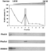
Subcellular localization of PhaZd determined by sucrose density gradient centrifugation. Ten microliters of each fraction (1.1 ml) was analyzed by Western blotting and immunostaining with antiserum against PhaZdWeu, PhaZa1Weu, or 3HB dehydrogenase (3HBDH). Columns and circles show the amount of PHB and protein, respectively.
FIG. 5.
FIG. 5.
Growth, PHB accumulation, and expression of PhaZd. A and C: dry cell weight (upper) and accumulation of PHB (lower). The wild-type and ΔphaZd mutant cells were cultured in nutrient-rich medium (A) or PHB-accumulating (C, solid line) or PHB-degrading (C, dotted line) conditions. PHB accumulation: cells were grown in minimal salt medium containing 2% (wt/vol) fructose and 0.1% (wt/vol) ammonium sulfate. PHB degradation; 60-h-cultured cells that had accumulated PHB were transferred to medium containing 0.5% (wt/vol) ammonium sulfate (arrows). Solid circle, wild type; open circle, H16DZd1 (ΔphaZd mutant). Results are the means for three independent measurements. The range of error bars was within ±5% of each value in the wild-type and ±10% in H16DZd1. B and D: time-dependent immunostaining analysis using the antiserum against PhaZdWeu. Asterisks show the time after the transfer. Each lane contained 50 μg of protein.
FIG. 6.
FIG. 6.
Comparison of structures of PhaZdWeu, PhaZ1Rru, and extracellular PHB depolymerases. Reversed characters (S, D, H) show amino acid residues in the catalytic triad. An asterisk indicates the histidine of the supposed oxyanion hole. SP, signal peptide; SBD, substrate binding domain.

Similar articles

Cited by

References

    1. Berg, J., J. Tymoczko, and L. Stryer. 2002. Biochemistry, 5th ed., p. 190-225. W. H. Freeman and Company, New York, N.Y.
    1. Bernhard, M., B. Benelli, A. Hochkoeppler, D. Zannoni, and B. Friedrich. 1997. Functional and structural role of the cytochrome b subunit of the membrane-bound hydrogenase complex of Alcaligenes eutrophus H16. Eur. J. Biochem. 248:179-186. - PubMed
    1. Bernhard, M., B. Friedrich, and R. A. Siddiqui. 2000. Ralstonia eutropha TF93 is blocked in tat-mediated protein export. J. Bacteriol. 182:581-588. - PMC - PubMed
    1. Cumming, R. C., N. L. Andon, P. A. Haynes, M. Park, W. H. Fischer, and D. Schubert. 2004. Protein disulfide bond formation in the cytoplasm during oxidative stress. J. Biol. Chem. 279:21749-21758. - PubMed
    1. Daws, E. A., and D. W. Ribbons. 1964. Some aspects of the endogenous metabolism of bacteria. Bacteriol. Rev. 28:126-149. - PMC - PubMed

Publication types

MeSH terms

Substances

LinkOut - more resources