. 2005 Nov;17(11):2911-21.
doi: 10.1105/tpc.105.035394. Epub 2005 Sep 30.

RNA interference identifies a calcium-dependent protein kinase involved in Medicago truncatula root development

Affiliations

RNA interference identifies a calcium-dependent protein kinase involved in Medicago truncatula root development

Sergey Ivashuta et al. Plant Cell. 2005 Nov.

Abstract

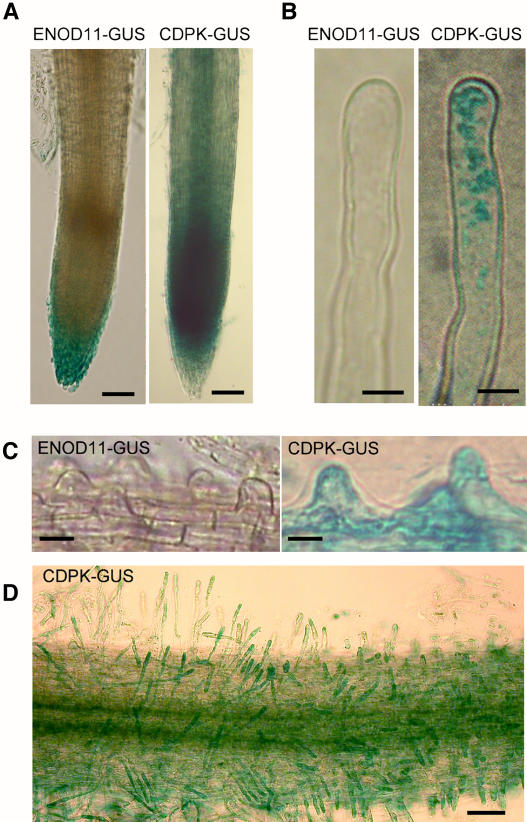
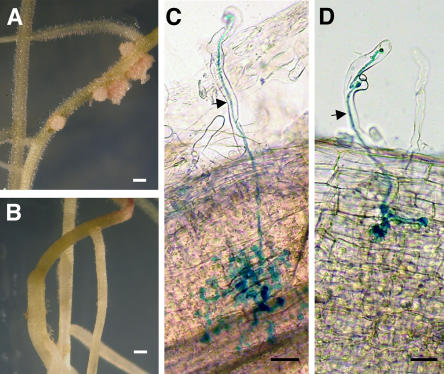
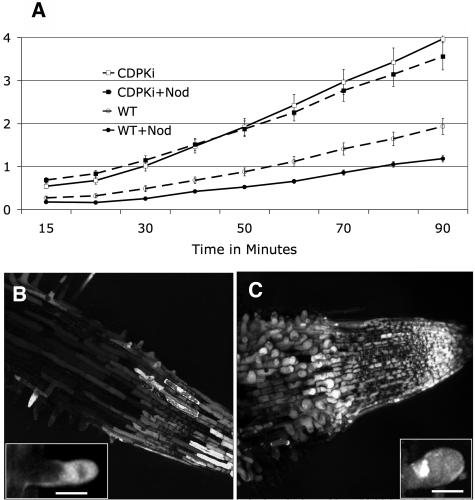
Changes in cellular or subcellular Ca2+ concentrations play essential roles in plant development and in the responses of plants to their environment. However, the mechanisms through which Ca2+ acts, the downstream signaling components, as well as the relationships among the various Ca2+-dependent processes remain largely unknown. Using an RNA interference-based screen for gene function in Medicago truncatula, we identified a gene that is involved in root development. Silencing Ca2+-dependent protein kinase1 (CDPK1), which is predicted to encode a Ca2+-dependent protein kinase, resulted in significantly reduced root hair and root cell lengths. Inactivation of CDPK1 is also associated with significant diminution of both rhizobial and mycorrhizal symbiotic colonization. Additionally, microarray analysis revealed that silencing CDPK1 alters cell wall and defense-related gene expression. We propose that M. truncatula CDPK1 is a key component of one or more signaling pathways that directly or indirectly modulates cell expansion or cell wall synthesis, possibly altering defense gene expression and symbiotic interactions.


Figures

Figure 1.
Figure 1.
Characterization of M. truncatula CDPK1-Silenced (CDPKi) Roots. Comparisons of control (left) and CDPKi (right) roots are shown in (A) to (D). (A) and (B) Suppression of CDPK1 gene expression results in short roots (A) and short root hairs (B). Bar = 100 μm. (C) RT-PCR analysis shows significant suppression of CDPK1 gene expression in CDPKi roots. Primers P2 (sense) and P3 (antisense) are within the region of the gene used for the pRNAi1444-1 construct, and primers P1 (sense) and P4 (antisense) are upstream and downstream of this region, respectively. (D) Root hair phenotypes of control and CDPKi. Bars = 50 μm. (E) Cortical root cell elongation is affected in CDPKi. Bars = 30 μm.
Figure 2.
Figure 2.
Roots Containing the PROCDPK1-GUS (CDPK-GUS) Reporter Gene Stained for GUS Expression. (A) to (C) A high level of promoter activity was detected in the root elongation zone (A) (bars = 150 μm) and in actively growing (B) and emerging (C) root hairs (bars = 10 μm) compared with the PROENOD11-GUS (ENOD11-GUS) reporter gene used as a control. (D) Portion of a root expressing the PROCDPK1-GUS reporter gene just above the root elongation zone. Bar = 80 μm.
Figure 3.
Figure 3.
Abnormalities in Root Hair Development Caused by Decreased Expression of CDPK1. (A) Fully elongated root hairs on CDPKi roots. Enlargements of representative root hairs are shown in separate panels at right. Bar = 30 μm. (B) Untreated (−Nod) and Nod factor–treated (+Nod) actively growing root hairs on control roots (see Methods). For Nod factor treatment, roots were incubated for 36 h with 10 nM Nod factor diluted in water. Bars = 30 μm.
Figure 4.
Figure 4.
Suppression of CDPK1 Gene Expression Resulted in Reduced Efficiency of Nodulation. (A) and (B) Control (A) and representative CDPKi (B) roots inoculated with S. meliloti and grown for 3 weeks on buffered nodulation medium supplemented with 1 mM α-aminoisobutyric acid, an ethylene inhibitor that promotes nodulation. Bars = 300 μm. (C) and (D) Control (C) and CDPKi (D) roots inoculated with S. meliloti carrying a LacZ reporter gene and grown on Turface and stained with X-Gal at 7 d after inoculation.
Figure 5.
Figure 5.
Mycorrhizal Colonization Is Diminished in CDPKi Roots. Control (left column) and CDPKi (right column) roots were inoculated with G. versiforme, harvested at 17 d after inoculation, and stained with WGA-Alexa Fluor 488 (Molecular Probes) to enable visualization of the fungus. Horizontal spread of intercellular hyphae through the cortex is reduced in CDPKi roots (top row) compared with control roots. An overlay of the bright-field and fluorescence micrographs is shown in the middle row. The bottom row shows close-up views of the infection sites. The arrowheads point to arbuscules, and the arrows point to intercellular hyphae. Bars = 50 μm.
Figure 6.
Figure 6.
Cell Wall Composition and Gene Expression Are Altered in CDPKi Roots. (A) Classification of genes whose expression was altered in CDPKi roots. Ratios of CDPKi to control root gene expression were obtained from microarray experiments. (B) CDPKi roots contain increased levels of lignin (pink coloration). Control and CDPKi plants were stained with phloroglucinol, and representative roots are shown. Bar = 100 μm. (C) Micrographs of root sections taken under UV light to visualize cell wall autofluorescence. Arrows point to vascular bundles. Bars = 25 μm.
Figure 7.
Figure 7.
Expression of CDPK1 in Different Plant Organs and in Response to Various Treatments. (A) CDPK1 expressed at similar levels in various plant organs. Seven micrograms of total RNA from flowers (F), stems (S), leaves (L), and roots (R) was used for RNA gel blot analysis. (B) Histogram showing levels of CDPK1 expression in response to treatment of roots with 2,4-D (50 nM), 1-aminocyclopropane-1-carboxylic acid (ACC; 5 μM), and abscisic acid (ABA; 10 μM) for 24 h; wounding (30 min); desiccation (∼30% of weight loss); and inoculation with S. meliloti (1, 3, and 48 h). Results of the RNA gel blot analyses were normalized to nontreated controls used in each experiment, and the ratio of expression in treated versus control roots is presented. NIH Image software (Scion) was used to analyze band intensity. (C) Activation of PROCDPK1-GUS reporter gene expression in roots by wounding.
Figure 8.
Figure 8.
ROS Accumulation Is Altered in CDPKi Roots. (A) ROS measurement (arbitrary units; se is shown as vertical bars) using the Amplex red hydrogen peroxide/peroxidase assay kit in 1-cm-long roots. (B) and (C) Confocal microscopy images of roots stained for ROS accumulation (CM-H2DCF imaging). The root hair initiation zone and root elongation zone of control (B) and CDPKi (C) are shown. Insets show representative root hairs. Bars = 20 μm.
Figure 9.
Figure 9.
The Actin Cytoskeleton of Root Cells Visualized with Alexa Fluor 488 Phalloidin. (A) and (B) Control root hair tip (A) and mid region (B). Arrows point to large bundles of actin filaments. (C) and (D) Representative CDPKi root hairs with an extreme length phenotype. The bright fluorescence foci suggest the accumulation of short microfilament fragments (arrowheads). (E) and (F) Control (E) and CDPKi (F) root epidermal cells. Bars = 10 μm.

References

    1. Allwood, E.G., Smertenko, A.P., and Hussey, P.J. (2001). Phosphorylation of plant actin depolymerising factor by calmodulin-like domain protein kinase. FEBS Lett. 499, 97–100. - PubMed
    1. Boisson-Dernier, A., Chabaud, M., Garcia, F., Becard, G., Rosenberg, C., and Barker, D.G. (2001). Agrobacterium rhizogenes-transformed roots of Medicago truncatula for the study of nitrogen-fixing and endomycorrhizal symbiotic associations. Mol. Plant Microbe Interact. 14, 695–700. - PubMed
    1. Brewin, N.J. (2004). Plant cell wall remodelling in the rhizobium–legume symbiosis. CRC Crit. Rev. Plant Sci. 23, 293–316.
    1. Cano-Delgado, A., Penfield, S., Smith, C., Catley, M., and Bevan, M. (2003). Reduced cellulose synthesis invokes lignification and defense responses in Arabidopsis thaliana. Plant J. 34, 351–362. - PubMed
    1. Catoira, R., Galera, C., de Billy, F., Penmetsa, R.V., Journet, E.P., Maillet, F., Rosenberg, C., Cook, D., Gough, C., and Denarie, J. (2000). Four genes of Medicago truncatula controlling components of a nod factor transduction pathway. Plant Cell 12, 1647–1666. - PMC - PubMed

Publication types

MeSH terms

LinkOut - more resources