Localization of Ptr ToxA Produced by Pyrenophora tritici-repentis Reveals Protein Import into Wheat Mesophyll Cells
- PMID: 16199615
- PMCID: PMC1276038
- DOI: 10.1105/tpc.105.035063
Localization of Ptr ToxA Produced by Pyrenophora tritici-repentis Reveals Protein Import into Wheat Mesophyll Cells
Abstract
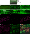
The plant pathogenic fungus Pyrenophora tritici-repentis secretes host-selective toxins (HSTs) that function as pathogenicity factors. Unlike most HSTs that are products of enzymatic pathways, at least two toxins produced by P. tritici-repentis are proteins and, thus, products of single genes. Sensitivity to these toxins in the host is conferred by a single gene for each toxin. To study the site of action of Ptr ToxA (ToxA), toxin-sensitive and -insensitive wheat (Triticum aestivum) cultivars were treated with ToxA followed by proteinase K. ToxA was resistant to protease, but only in sensitive leaves, suggesting that ToxA is either protected from the protease by association with a receptor or internalized. Immunolocalization and green fluorescent protein tagged ToxA localization demonstrate that ToxA is internalized in sensitive wheat cultivars only. Once internalized, ToxA localizes to cytoplasmic compartments and to chloroplasts. Intracellular expression of ToxA by biolistic bombardment into both toxin-sensitive and -insensitive cells results in cell death, suggesting that the ToxA internal site of action is present in both cell types. However, because ToxA is internalized only in sensitive cultivars, toxin sensitivity, and therefore the ToxA sensitivity gene, are most likely related to protein import. The results of this study show that the ToxA protein is capable of crossing the plant plasma membrane from the apoplastic space to the interior of the plant cell in the absence of a pathogen.
Figures


Similar articles
-
Ptr ToxA interacts with a chloroplast-localized protein.Mol Plant Microbe Interact. 2007 Feb;20(2):168-77. doi: 10.1094/MPMI-20-2-0168. Mol Plant Microbe Interact. 2007. PMID: 17313168
-
Structure of Ptr ToxA: an RGD-containing host-selective toxin from Pyrenophora tritici-repentis.Plant Cell. 2005 Nov;17(11):3190-202. doi: 10.1105/tpc.105.034918. Epub 2005 Oct 7. Plant Cell. 2005. PMID: 16214901 Free PMC article.
-
Emergence of tan spot disease caused by toxigenic Pyrenophora tritici-repentis in Australia is not associated with increased deployment of toxin-sensitive cultivars.Phytopathology. 2008 May;98(5):488-91. doi: 10.1094/PHYTO-98-5-0488. Phytopathology. 2008. PMID: 18943215
-
Host-selective toxins, Ptr ToxA and Ptr ToxB, as necrotrophic effectors in the Pyrenophora tritici-repentis-wheat interaction.New Phytol. 2010 Sep;187(4):911-9. doi: 10.1111/j.1469-8137.2010.03362.x. Epub 2010 Jul 14. New Phytol. 2010. PMID: 20646221 Review.
-
Host-specific toxins: effectors of necrotrophic pathogenicity.Cell Microbiol. 2008 Jul;10(7):1421-8. doi: 10.1111/j.1462-5822.2008.01153.x. Epub 2008 Apr 1. Cell Microbiol. 2008. PMID: 18384660 Review.
Cited by
-
Proteome changes induced by Pyrenophora tritici-repentis ToxA in both insensitive and sensitive wheat indicate senescence-like signaling.Proteome Sci. 2015 Feb 5;13:3. doi: 10.1186/s12953-014-0060-3. eCollection 2015. Proteome Sci. 2015. PMID: 25663824 Free PMC article.
-
Genetic characterization of adult-plant resistance to tan spot (syn, yellow spot) in wheat.Theor Appl Genet. 2021 Sep;134(9):2823-2839. doi: 10.1007/s00122-021-03861-8. Epub 2021 Jun 1. Theor Appl Genet. 2021. PMID: 34061222
-
Phytotoxicity and innate immune responses induced by Nep1-like proteins.Plant Cell. 2006 Dec;18(12):3721-44. doi: 10.1105/tpc.106.044180. Epub 2006 Dec 28. Plant Cell. 2006. PMID: 17194768 Free PMC article.
-
A single binding site mediates resistance- and disease-associated activities of the effector protein NIP1 from the barley pathogen Rhynchosporium secalis.Plant Physiol. 2007 Jul;144(3):1654-66. doi: 10.1104/pp.106.094912. Epub 2007 May 3. Plant Physiol. 2007. PMID: 17478637 Free PMC article.
-
The essential effector SCRE1 in Ustilaginoidea virens suppresses rice immunity via a small peptide region.Mol Plant Pathol. 2020 Apr;21(4):445-459. doi: 10.1111/mpp.12894. Epub 2020 Feb 22. Mol Plant Pathol. 2020. PMID: 32087618 Free PMC article.
References
-
- Alfano, J.R., and Collmer, A. (2004). Type III secretion system effector proteins: Double agents in bacterial disease and plant defense. Annu. Rev. Phytopathol. 42, 385–414. - PubMed
-
- Allen, R.L., Bittner-Eddy, P.D., Grenville-Briggs, L.J., Meitz, J.C., Rehmany, A.P., Rose, L.E., and Beynon, J.L. (2004). Host-parasite coevolutionary conflict between Arabidopsis and downy mildew. Science 306, 1957–1960. - PubMed
-
- Anderson, J.A., Effertz, R.J., Faris, J.D., Francl, L.J., Meinhardt, S.W., and Gill, B.S. (1999). Genetic analysis of sensitivity to a Pyrenophora tritici-repentis necrosis-inducing toxin in durum and common wheat. Phytopathology 89, 293–297. - PubMed
-
- Ballance, G.M., Lamari, L., and Bernier, C.C. (1989). Purification and characterization of a host-selective necrosis toxin from Pyrenophora tritici-repentis. Physiol. Mol. Plant Pathol. 35, 203–213.
Publication types
MeSH terms
Substances
Grants and funding
LinkOut - more resources
Full Text Sources
Medical

