

Structure of a shark IgNAR antibody variable domain and modeling of an early-developmental isotype
- PMID: 16199666
- PMCID: PMC2253229
- DOI: 10.1110/ps.051709505
Structure of a shark IgNAR antibody variable domain and modeling of an early-developmental isotype
Abstract
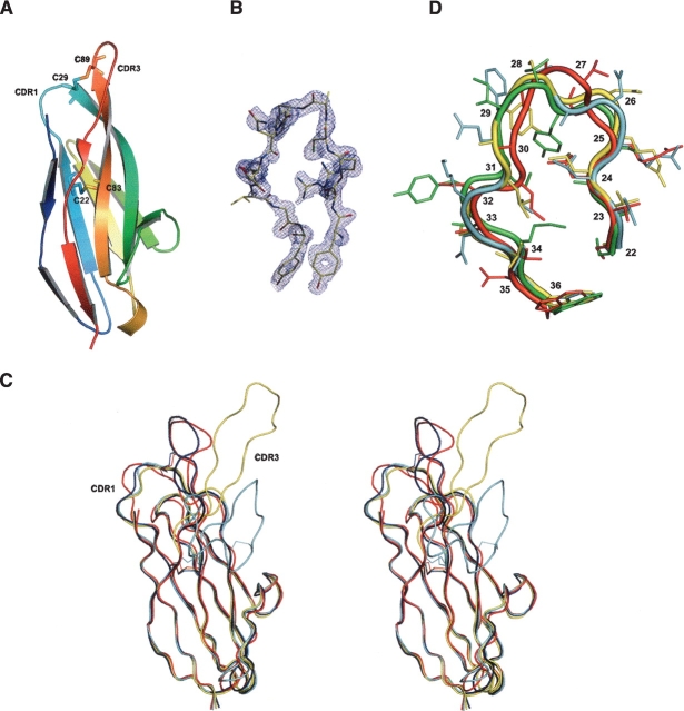
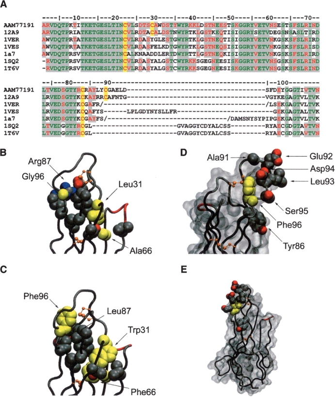
The new antigen receptor (IgNAR) antibodies from sharks are disulphide bonded dimers of two protein chains, each containing one variable and five constant domains. Three types of IgNAR variable domains have been discovered, with Type 3 appearing early in shark development and being overtaken by the antigen-driven affinity-matured Type 1 and 2 response. Here, we have determined the first structure of a naturally occurring Type 2 IgNAR variable domain, and identified the disulphide bond that links and stabilizes the CDR1 and CDR3 loops. This disulphide bridge locks the CDR3 loop in an "upright" conformation in contrast to other shark antibody structures, where a more lateral configuration is observed. Further, we sought to model the Type 3 isotype based on the crystallographic structure reported here. This modeling indicates (1) that internal Type 3-specific residues combine to pack into a compact immunoglobulin core that supports the CDR loop regions, and (2) that despite apparent low-sequence variability, there is sufficient plasticity in the CDR3 loop to form a conformationally diverse antigen-binding surface.
Figures
Similar articles
-
Maturation of shark single-domain (IgNAR) antibodies: evidence for induced-fit binding.J Mol Biol. 2007 Mar 23;367(2):358-72. doi: 10.1016/j.jmb.2006.12.045. Epub 2006 Dec 22. J Mol Biol. 2007. PMID: 17258766
-
Isolation and characterization of an IgNAR variable domain specific for the human mitochondrial translocase receptor Tom70.Eur J Biochem. 2003 Sep;270(17):3543-54. doi: 10.1046/j.1432-1033.2003.03737.x. Eur J Biochem. 2003. PMID: 12919318
-
Rapid isolation of IgNAR variable single-domain antibody fragments from a shark synthetic library.Mol Immunol. 2007 Jan;44(4):656-65. doi: 10.1016/j.molimm.2006.01.010. Epub 2006 Feb 24. Mol Immunol. 2007. PMID: 16500706
-
Shark New Antigen Receptor (IgNAR): Structure, Characteristics and Potential Biomedical Applications.Cells. 2021 May 8;10(5):1140. doi: 10.3390/cells10051140. Cells. 2021. PMID: 34066890 Free PMC article. Review.
-
Incorporation of long CDR3s into V domains: implications for the structural evolution of the antibody-combining site.Exp Clin Immunogenet. 2001;18(4):176-98. doi: 10.1159/000049197. Exp Clin Immunogenet. 2001. PMID: 11872949 Review.
Cited by
-
The immunoglobulins of cartilaginous fishes.Dev Comp Immunol. 2021 Feb;115:103873. doi: 10.1016/j.dci.2020.103873. Epub 2020 Sep 23. Dev Comp Immunol. 2021. PMID: 32979434 Free PMC article. Review.
-
Allosteric inhibition of Aurora-A kinase by a synthetic vNAR domain.Open Biol. 2016 Jul;6(7):160089. doi: 10.1098/rsob.160089. Open Biol. 2016. PMID: 27411893 Free PMC article.
-
Screening and characterization of inhibitory vNAR targeting nanodisc-assembled influenza M2 proteins.iScience. 2022 Dec 5;26(1):105736. doi: 10.1016/j.isci.2022.105736. eCollection 2023 Jan 20. iScience. 2022. PMID: 36570769 Free PMC article.
-
vNARs as Neutralizing Intracellular Therapeutic Agents: Glioblastoma as a Target.Antibodies (Basel). 2024 Mar 18;13(1):25. doi: 10.3390/antib13010025. Antibodies (Basel). 2024. PMID: 38534215 Free PMC article. Review.
-
Hepatitis B precore protein: pathogenic potential and therapeutic promise.Yonsei Med J. 2012 Sep;53(5):875-85. doi: 10.3349/ymj.2012.53.5.875. Yonsei Med J. 2012. PMID: 22869468 Free PMC article. Review.
References
-
- Adelman, M.K., Schluter, S.F., and Marchalonis, J.J. 2004. The natural antibody repertoire of sharks and humans recognizes the potential universe of antigens. Protein J. 23 103–118. - PubMed
-
- Al-Lazikani, B., Lesk, A.M., and Chothia, C. 1997. Standard conformations for the canonical structures of immunoglobulins. J. Mol. Biol. 273 927–948. - PubMed
-
- Brünger, A.T., Adams, P.D., Clore, G.M., DeLano, W.L., Gros, P., Grosse-Kunstleve, R.W., Jiang, J.S., Kuszewski, J., Nilges, M., Pannu, N.S., et al. 1998. Crystallography & NMR system: A new software suite for macromolecular structure determination. Acta Crystallogr. D Biol. Crystallogr. 54 905–921. - PubMed
-
- Chothia, C., Lesk, A.M., Tramontano, A., Levitt, M., Smith-Gill, S.J., Air, G., Sheriff, S., Padlan, E.A., Davies, D., Tulip, W.R., et al. 1989. Conformations of immunoglobulin hypervariable regions. Nature 28 877–883. - PubMed
-
- Coia, G., Ayres, A., Lilley, G.G., Hudson, P.J., and Irving, R.A. 1997. Use of mutator cells as a means for increasing production levels of a recombinant antibody directed against Hepatitis B. Gene 201 203–209. - PubMed
Publication types
MeSH terms
Substances
LinkOut - more resources
Full Text Sources
Other Literature Sources
