. 2005 Oct;25(20):8913-24.
doi: 10.1128/MCB.25.20.8913-8924.2005.

The amino-terminal region of Drosophila MSL1 contains basic, glycine-rich, and leucine zipper-like motifs that promote X chromosome binding, self-association, and MSL2 binding, respectively

Affiliations

The amino-terminal region of Drosophila MSL1 contains basic, glycine-rich, and leucine zipper-like motifs that promote X chromosome binding, self-association, and MSL2 binding, respectively

Fang Li et al. Mol Cell Biol. 2005 Oct.

Abstract

In Drosophila melanogaster, X chromosome dosage compensation is achieved by doubling the transcription of most X-linked genes. The male-specific lethal (MSL) complex is required for this process and binds to hundreds of sites on the male X chromosome. The MSL1 protein is essential for X chromosome binding and serves as a central scaffold for MSL complex assembly. We find that the amino-terminal region of MSL1 binds to hundreds of sites on the X chromosome in normal males but only to approximately 30 high-affinity sites in the absence of endogenous MSL1. Binding to the high-affinity sites requires a basic motif at the amino terminus that is conserved among Drosophila species. X chromosome binding also requires a conserved leucine zipper-like motif that binds to MSL2. A glycine-rich motif between the basic and leucine-zipper-like motifs mediates MSL1 self-association in vitro and binding of the amino-terminal region of MSL1 to the MSL complex assembled on the male X chromosome. We propose that the basic region may mediate DNA binding and that the glycine-rich region may promote the association of MSL complexes to closely adjacent sites on the X chromosome.


Figures
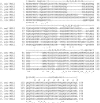
FIG. 1.
FIG. 1.
Alignment of the amino-terminal domain sequences of MSL1 homologs from seven Drosophila species. Likely homologs of D. melanogaster MSL1 were identified from the completed genome sequence of D. pseudoobscura (D. pse) and the draft genome sequences of D. simulans (D. sim), D. yakuba (D. yak), D. erecta (D. ere), and D. virilis (D. vir). The basic region (aa 1 to 15), apolar region (aa 113 to 121), glutamine (Q)-rich region (aa 122 to 127), and coiled-coil domain (aa 128 to 159) are all well conserved. While few individual amino acids in the glycine (G)-, proline (P)-, histidine (H)-, and asparagine (N)-rich domain (aa 16 to 112) are well conserved, all Drosophila MSL1s contain a high proportion of G, P, H, and N in this region.
FIG. 2.
FIG. 2.
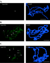
MSL1NHA binds to hundreds of sites on the male X chromosome. (A) Schematic representation of the msl1 constructs used to make transgenic flies. All constructs carry an HA epitope tag at the end of the open reading frame. Expression was controlled using the heat-inducible hsp70 promoter. The boxed region indicates the apolar, Q-rich, and coiled-coil regions highlighted in Fig. 1. For the alanine replacement mutations, the amino acids that are changed are indicated in parentheses. (B) Western blot with anti-HA antibody and protein extracts from adult flies. Protein of the anticipated mass is detected in extracts from all lines. (C to F) MSL1NHA but not Δ84HA binds to the male X chromosome. Male salivary gland nuclei were stained with anti-HA antibody (green) and counterstained with DAPI (blue) to visualize all of the chromosomes. The hsp70 promoter was used to control MSL1NHA and Δ84HA gene expression in all lines except the results shown in panel E, where the constitutive armadillo promoter (45) was used. All larvae were raised at 25°C and not heat shocked with the exception of the results shown in panel C, where larvae were treated at 37°C for 20 min and then left to recover at 25°C for 4 h. MSL1NHA was detected at hundreds of sites on the male X chromosome either with (C) or without (D) heat shock and with either promoter (C and E). In contrast, no binding of Δ84HA was detected to the male X chromosome (arrowhead) in both heat-treated (not shown) and unshocked (F) larvae. (G to I) Both MSL1NHA and Δ84HA are localized to the nucleus. Whole male salivary glands with intact nuclei were stained with anti-HA antibody (green) and counterstained with DAPI (blue). MSL1NHA (G) and Δ84HA (H and I) are detected in the nucleus. No nuclear staining was detected in the wild-type untransformed control (J). Scale bar, either 30 μm (C to F) or 10 μm (G to J).
FIG. 3.
FIG. 3.
MSL1HA binds to ∼30 high-affinity sites on the X chromosome in larvae that lack endogenous MSL1. (A) Homozygous msl1L60 female salivary gland nuclei that express MSL2 (hsp83-msl2) and MSL1NHA (hsp70 promoter) were stained with anti-HA (green), anti-MSL2 (red) (top), or anti-MOF (red) (bottom) and counterstained with DAPI (blue). The high-affinity sites at 3F, 10C, and 17F are indicated. MSL2 but not MOF colocalizes with MSL1NHA to ∼30 high-affinity sites in larvae that are homozygous for msl1L60, a ∼2.5-kb deletion of most of the msl1 gene (R. Kelley, personal communication). (B) MSL1NHA binds to hundreds of sites on the X chromosome in female larvae that express MSL2 and are heterozygous for a null mutation in msl1. Heterozygous msl1L60 female salivary gland nuclei that express MSL2 and MSL1NHA were stained with the antibodies, as in the results shown in panel A. Scale bar, 30 μm.
FIG. 4.
FIG. 4.
Binding pattern of MSL1 amino-terminal deletion mutants to the male X chromosome. (A to C) Male salivary gland nuclei were stained with anti-HA antibody (green) and anti-MSL2 (red) and counterstained with DAPI (blue). Δ74HA does not bind to X chromosome (A), Δ50HA binds weakly (B), and Δ26HA binds more strongly to the X chromosome than Δ50HA (C); but staining intensity is consistently less than for MSL1NHA (Fig. 2C and D). Scale bar, 30 μm.
FIG. 5.
FIG. 5.
The amino-terminal basic region is essential for binding to the X chromosome in the absence of endogenous MSL1. (A) Δ26HA does not bind to the X chromosome (arrowhead). Homozygous msl1L60 female salivary gland nuclei that express MSL2 (hsp83-msl2) and Δ26HA were stained with anti-HA (green) and counterstained with DAPI (blue). (B) Replacement by alanine of three conserved basic amino acids in the amino-terminal basic motif eliminates binding to most of the high-affinity sites. Homozygous msl1L60 female salivary gland nuclei that express MSL2 (hsp83-msl2) and mut_bas1 were stained with anti-HA (green) and counterstained with DAPI (blue). Binding was detected only to the high-affinity sites at 3F, 8F, 10C, 11B, and 17F as indicated. (C) Replacement by alanine of two conserved aromatic amino acids in the amino-terminal basic motif increases binding to the autosomes. Homozygous msl1L60 female salivary gland nuclei that express MSL2 (hsp83-msl2) and mut_bas2 were stained with anti-HA (green) and counterstained with DAPI (blue). mut_bas2 binds to all of the high-affinity sites on the X chromosome and also binds to more autosomal sites than MSL1NHA (arrowheads). Scale bar, 30 μm.
FIG. 6.
FIG. 6.
The glycine-rich region mediates MSL1 self-association. Protein extracts from transformant flies that cooverexpressed an HA-tagged MSL1 amino-terminal domain and either MSL1 (A), MSL3 (B), or MSL2 (C) were immunoprecipitated with anti-HA affinity matrix (IP) and detected by Western blotting (IB) with indicated antibodies. Immunoprecipitated extracts (Ip) are shown in the even numbered lanes and 10% of the corresponding input is shown in the odd-numbered lanes (Input). (A) MSL1 coimmunoprecipitates with MSL1NHA but not Δ84HA. Protein extracts were from flies that cooverexpressed MSL1; either MSL1NHA (lanes 1 and 2), Δ84HA (lanes 3 and 4), Δ74HA (lanes 5 and 6), Δ50HA (lanes 7 and 8), or Δ26HA (lanes 9 and 10) was immunoprecipitated and detected by Western blotting with anti-MSL1. MSL1 (arrow) coimmunoprecipitated with MSL1NHA and Δ26HA but not with any of the other amino-terminal deletion mutants (arrows). (B) MSL3 does not coimmunoprecipitate with Δ26HA. Protein from flies that cooverexpressed MSL3 and Δ26HA (lanes 1 and 2) were immunoprecipitated and detected by Western blotting with either anti-MSL3 (top) or anti-HA (bottom). (C) MSL2 coimmunoprecipitates with both MSL1NHA and Δ84HA. Protein from flies that cooverexpressed MSL2 and either MSL1NHA (lanes 1 and 2) or Δ84HA (lanes 3 and 4) was immunoprecipitated and detected by Western blotting with either anti-MSL2 (top) or anti-HA (bottom). MSL2 (arrow) coimmunoprecipitates with both MSL1NHA and Δ84HA. (B and C) Western blotting with anti-HA antibody shows the efficiency of immunoprecipitation. Lower-molecular-weight bands were occasionally detected in immunoprecipitated protein (asterisk), which are presumably products of partial protein degradation.
FIG. 7.
FIG. 7.
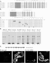
The leucine zipper-like motif of MSL1 is required for binding to MSL2. (A) Alignment of the coiled-coil regions of orthologs of D. melanogaster (Dme) MSL1 from the mosquito Anopheles gambiae (Aga), from the fish Takifugu rubripes (Tru), zebra fish Danio rerio (Dre), and human Homo sapiens (Hsa). The apolar, glutamine (Q)-rich, and leucine zipper-like regions are indicated. The a and d positions of the coiled-coil motif, which are usually occupied by apolar amino acids, are indicated. The alignment is slightly different than that published previously for MSL1 orthologs (29). (B) Alignment of the amino-terminal region of orthologs of D. melanogaster (Dme) MSL2 from Drosophila virilis (Dvi), the fish T. rubripes (Tru), and humans (Hsa). The apolar and coiled-coil regions are indicated. (C) Alanine substitution mutations in MSL1NHA used in this study. (D) Coimmunoprecipitation of the amino-terminal domains of MSL2 and MSL1. In vitro-translated [35S]methionine-labeled MSL1NHA, the carboxyl-terminal domain of MSL1 (aa 705 to 1039) (41), and alanine substitution derivatives of MSL1NHA were incubated with protein extracts from either wild-type flies or transgenic flies that had been prebound to anti-FLAG affinity beads. The transgenic flies overexpressed MSL2NFLAG, which is the amino-terminal domain of MSL2 (aa 1 to 193) with a FLAG tag at the C end. Bound proteins immunoprecipitated with anti-FLAG affinity beads, were separated by SDS-PAGE, and were detected by autoradiography. [35S]methionine-labeled proteins coimmunoprecipitated with MSL2NFLAG (p) are shown in lanes 2, 5, 7, 9, 11, 13, 15, and 17; those coimmunoprecipitated with extract from control wild-type flies (p-) are shown in lane 3; the other lanes had 10% of the corresponding input (i). MSL1NA (lane 2), mut_QEQ (lane 9), mut_cc3 (lane 15), and mut_cc4 (lane 17) all efficiently coimmunoprecipitated with MSL2NFLAG. MSL1NHA did not coimmunoprecipitate with the negative control extract from untransformed wild-type flies (lane 3). Significantly less coimmunoprecipitation was seen with mut_cc1 (lane 11) and mut_cc2 (lane 13). MSL1C (lane 5) and mut_apo (lane 7) did not coimmunoprecipitate with MSL2NFLAG. (E) Western blot of coimmunoprecipitated samples with anti-FLAG antibody. Equivalent aliquots of immunoprecipitated protein used in the results shown in panel D were size separated by SDS-PAGE, and MSL2NFLAG was detected by Western blotting. The lane numbers correspond to the identical samples used above (D). (F and G) mut_QEQ binds to the male X chromosome but mut_apo does not. Male salivary gland nuclei that express either mut_apo (F) or mut_QEQ (G) were stained with anti-HA antibody and counterstained with DAPI. The arrowhead (F) points to the male X chromosome. Scale bar, 30 μm.

Similar articles

Cited by

References

    1. Akhtar, A. 2003. Dosage compensation: an intertwined world of RNA and chromatin remodelling. Curr. Opin. Genet. Dev. 13:161-169. - PubMed
    1. Akhtar, A., and P. B. Becker. 2000. Activation of transcription through histone H4 acetylation by MOF, an acetyltransferase essential for dosage compensation in Drosophila. Mol. Cell 5:367-375. - PubMed
    1. Akhtar, A., D. Zink, and P. B. Becker. 2000. Chromodomains are protein-RNA interaction modules. Nature 407:405-409. - PubMed
    1. Bashaw, G. J., and B. S. Baker. 1995. The msl-2 dosage compensation gene of Drosophila encodes a putative DNA-binding protein whose expression is sex specifically regulated by Sex-lethal. Development 121:3245-3258. - PubMed
    1. Belote, J. M., and J. C. Lucchesi. 1980. Control of X chromosome transcription by the maleless gene in Drosophila. Nature 285:573-575. - PubMed

Publication types

MeSH terms

LinkOut - more resources