. 2005 Oct;25(20):8948-59.
doi: 10.1128/MCB.25.20.8948-8959.2005.

Ablation of MEKK4 kinase activity causes neurulation and skeletal patterning defects in the mouse embryo

Affiliations

Ablation of MEKK4 kinase activity causes neurulation and skeletal patterning defects in the mouse embryo

Amy N Abell et al. Mol Cell Biol. 2005 Oct.

Abstract

Skeletal disorders and neural tube closure defects represent clinically significant human malformations. The signaling networks regulating normal skeletal patterning and neurulation are largely unknown. Targeted mutation of the active site lysine of MEK kinase 4 (MEKK4) produces a kinase-inactive MEKK4 protein (MEKK4(K1361R)). Embryos homozygous for this mutation die at birth as a result of skeletal malformations and neural tube defects. Hindbrains of exencephalic MEKK4(K1361R) embryos show a striking increase in neuroepithelial cell apoptosis and a dramatic loss of phosphorylation of MKK3 and -6, mitogen-activated protein kinase kinases (MKKs) regulated by MEKK4 in the p38 pathway. Phosphorylation of MAPK-activated protein kinase 2, a p38 substrate, is also inhibited, demonstrating a loss of p38 activity in MEKK4(K1361R) embryos. In contrast, the MEK1/2-extracellular signal-regulated kinase 1 (ERK1)/ERK2 and MKK4-Jun N-terminal protein kinase pathways were unaffected. The p38 pathway has been shown to regulate the phosphorylation and expression of the small heat shock protein HSP27. Compared to the wild type, MEKK4(K1361R) fibroblasts showed significantly reduced phosphorylation of p38 and HSP27, with a corresponding heat shock-induced instability of the actin cytoskeleton. Together, these data demonstrate MEKK4 regulation of p38 and that substrates downstream of p38 control cellular homeostasis. The findings are the first demonstration that MEKK4-regulated p38 activity is critical for neurulation.


Figures
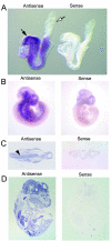
FIG. 1.
FIG. 1.
MEKK4 expression during embryonic development visualized by in situ hybridization with a RNA probe to the N-terminal half of MEKK4. (A) Whole-mount embryo in situ hybridization for MEKK4 at E8.5. The solid arrow indicates neural folds, and the open arrow indicates the allantois. (B) Whole-embryo in situ hybridization for MEKK4 at E10.5. (C) In situ hybridization for MEKK4 in cryosections isolated at E10.5. The arrowhead denotes neuroepithelium. (D) In situ hybridization for MEKK4 in cryosections of embryos isolated at E15.5.
FIG. 2.
FIG. 2.
Targeted replacement of the MEKK4 gene with a full-length kinase-inactive MEKK4K1361R. (A) Schematic diagram of MEKK4 gene, the targeting vector, and the targeted allele. Two mutations were generated in exon 21. One silent mutation engineered a new SacI restriction site in exon 21, and the second mutation produced the replacement of the active-site lysine at position 1361 by arginine, resulting in a full-length inactive kinase. The neomycin resistance positive selection marker was inserted in the reverse orientation in intron 21. The location of the hybridization probe is indicated. Digestion of genomic DNA with SacI produces the indicated fragments. (B) Mutation of the active site lysine of MEKK4 (MEKK4K1361R) disrupts the ability of MEKK4 to phosphorylate its substrate MKK6. 293 cells transfected with the HA-MEKK4wt kinase domain or HA-MEKK4K1361R kinase domain were immunoprecipitated with anti-HA antibody and incubated with purified kinase-inactive His-MKK6. Phosphorylation of His-MKK6 was detected with anti-phospho-MKK3/6 antibody. The total HA-MEKK4 kinase domain was detected with an anti-HA antibody. To show equal loading of substrate, Coomassie staining of the His-MKK6 substrate is shown. (C) Mutation of the active-site lysine of MEKK4 (MEKK4K1361R) disrupts the ability of MEKK4 to phosphorylate its substrate MKK4. 293 cells were transfected and immunoprecipitated as described in the legend to panel C and then incubated with inactive GST-MKK4. Phosphorylation of GST-MKK4 was detected with anti-phospho-MKK4 antibody. The total HA-MEKK4 kinase domain was detected with an anti-HA antibody. To show equal loading, the GST-MKK4 substrate was stained with Ponceau. (D) MEKK4K1361R is unable to activate p38. 293 cells were cotransfected with Flag-p38 and either Flag-MEKK4wt or Flag-MEKK4K1361R, and blots were probed as indicated. (E) Southern blot analysis of SacI-digested genomic DNA from two independent ES cell lines that were injected into C57BL/6 blastocysts and used to generate two independent heterozygous MEKK4K1361R mouse lines. (F) PCR analysis of genomic DNA isolated from tail clips from 3-week-old pups with primers in exons 21 and 22. (G) Western blot analysis of MEKK4 protein expression in lysates prepared from mouse embryo fibroblasts generated from littermate E14.5 embryos.
FIG. 3.
FIG. 3.
Skeletal defects in mice expressing kinase-inactive MEKK4K1361R. Bone (Alizarin red) and cartilage (Alcian blue) staining of ribcages from wild-type (A), homozygous knock-in of MEKK4K1361R (B), and heterozygote (C) fetuses isolated at E18.5, showing asymmetric attachment of sternocostal connections. (D) Quantitation of the frequency of rib misalignment in fetuses from heterozygote crosses. Dorsal views of whole animal bone and cartilage staining in wild-type (E and G) and a heterozygous knock-in of MEKK4K1361R (F and H), showing severe curvature of the spine, are shown. Magnification of vertebral columns of wild-type (G) and heterozygous mutant (H) fetuses, showing misalignment of vertebra in the mutant fetus. X-ray films showing scoliosis (J) and an abnormal tail (K) in homozygous knock-in MEKK4K1361R adult mice are compared to the wild type (I). Black arrowheads (E and F) indicate the area of vertebral column seen in the enlarged insets (G and H); the white arrowhead (J) indicates scoliosis; the white arrow (K) indicates bone remodeling in the tail.
FIG. 4.
FIG. 4.
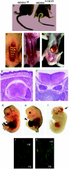
Neonates homozygous for MEKK4K1361R die at birth due to severe open neural tube and spine defects. (A) Posterior view of a normal adult wild-type mouse and the curly tail-spina bifida phenotype in a mouse homozygous for kinase-inactive MEKK4K1361R. (B) Dorsal view of dead MEKK4K1361R neonate, showing open spine. (C) Dorsal view of dead homozygous MEKK4K1361R neonate, showing rachischisis. (D) Ventral view of dead neonate homozygous for MEKK4K1361R, showing omphalocele. (E and F) Transverse sections taken from paraffin-embedded E18.5 fetuses stained with hematoxylin-eosin. (E) Normal spinal chord in heterozygote littermate control. (F) Open spinal chord in MEKK4K1361R fetus with spina bifida. (G to I) Embryos isolated at E14.5. Normal wild-type embryo (G) and mutant heterozygote embryos with omphalocele (H) or exencephaly (I). The arrowhead (H) indicates omphalocele and the arrow (I) indicates exencephaly. (J and K) TUNEL-labeled 10-μm cryosections from littermate E9.5 embryos. Apoptotic cells are shown in green. (J) The forebrain (FB) and hindbrain (HB) are closed in the wild-type embryo. (K) The hindbrain (HB) remains open and apoptosis is increased in a MEKK4K1361R mutant.
FIG. 5.
FIG. 5.
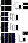
Inhibition of MKK3/6 and MAPKAPK-2 phosphorylation in MEKK4K1361R hindbrains. Cryosections from wild-type (A to C, H, I, M, N, and R to U) and homozygous MEKK4K1361R (D to F, J, K, O, P, and V to Y) E9.5 embryos isolated from timed heterozygote matings. (A and D) Phosphorylation of MKK3/6 is decreased in the hindbrains of MEKK4K1361R E9.5 embryos relative to wild-type littermates. A phospho-specific antibody for MKK3/6 was used to immunostain sections. (AA and DD) Enlarged insets of panels A and D, showing decreased phospho-MKK3/6 in MEKK4K1361R hindbrain. (B and E) TUNEL staining of sections showing enhanced apoptosis in the hindbrain of the MEKK4K1361R embryo. (C and F) Overlay of the phospho-MKK3/6 and TUNEL staining, showing the exclusion of apoptotic cells from areas with high phospho-MKK3/6 immunostaining. Phospho-MKK3/6 staining is in red, and TUNEL staining is in green. (G) Quantitation of mean phospho-MKK3/6 fluorescence intensity in the hindbrain. Data represent the means ± standard error of the mean (SEM) of three samples, each with P values of <0.001, indicating that differences are statistically significant. (H to K) Phosphorylation of MKK4 is similar in wild-type and mutant hindbrains. Phosphorylation of MKK4, shown in red, was detected with a phospho-specific antibody to MKK4; nuclei stained with DAPI are in blue. (L) Quantitation of mean phospho-MKK4 fluorescence intensity in the hindbrain. Data represent the means ± SEM of six samples, each with P values of >0.25, indicating that differences are not statistically significant. (M to P) Phosphorylation of MEK1/2 is similar in wild-type and mutant hindbrains. Phosphorylation of MEK1/2, shown in red, was detected with a phospho-specific antibody to MEK1/2; nuclei are shown in blue. (Q) Quantitation of mean phospho-MEK1/2 fluorescence intensity in the hindbrain. Data represent the means ± SEM of nine wild-type and five mutant samples with P values of >0.15, indicating that differences are not statistically significant. (R to Y) Phosphorylation of MAPKAPK-2 is decreased in the hindbrains of MEKK4K1361R E9.5 embryos relative to that of wild-type littermates. A phospho-specific antibody for MAPKAPK-2, shown in red, was used to immunostain sections; nuclei are shown in blue. (Z) Quantitation of mean phospho-MAPKAPK-2 fluorescence intensity in hindbrain. Data represent the means ± SEM of nine wild-type and eight mutant samples with P values of <0.001, indicating that differences are statistically significant. (RR) Enlarged inset of R, showing localization of phospho-MAPKAPK-2 at the site of neural tube closure in the wild-type hindbrain. Forebrain (FB) and hindbrain (HB) are closed in the wild-type embryo. The hindbrain remains open in the MEKK4K1361R mutants. *, P < 0.001. The white arrow indicates the site of hindbrain closure.
FIG. 6.
FIG. 6.
Inhibition of the p38 pathway in MEKK4K1361R MEFs. (A) Phosphorylation of p38 is inhibited in immortalized cycling homozygous and heterozygous MEFs generated from littermate E14.5 embryos. Phosphorylation of p38 (B) and MKK3 (C) in MEFs generated from primary cycling homozygous MEKK4K1361R embryos and littermate wild-type controls in cycling cells in response to TNF-α. MEFs were stimulated for the indicated times, and 15 μg of lysate was probed with total and phospho-specific antibodies to MKK3, MKK4, p38, and actin. (D) Phosphorylation of MKK3, p38, MAPKAPK-2, and Hsp27 is inhibited in MEKK4K1361R primary cycling MEFs. A total of 15 μg of lysate was probed with total and phospho-specific antibodies. The quantitation represents densitometry in relative units compared to the densitometry of actin for each sample. (E) Phosphorylation of p38 in response to heat shock is inhibited in MEKK4K1361R MEFs. Primary cycling MEFs were incubated at 42°C for the indicated times. A total of 25 μg of lysate was probed with the indicated total and phospho-specific antibodies. (F) Phosphorylation of HSP27 in response to heat shock is inhibited in MEKK4K1361R MEFs. Primary cycling MEFs were incubated at 42°C for the indicated times. A total of 25 μg of lysate was probed with the indicated total and phospho-specific antibodies.
FIG. 7.
FIG. 7.
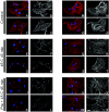
Increased thermosensitivity of actin microfilaments in primary MEKK4K1361R fibroblasts. Primary fibroblasts from littermate MEKK4K1361R (A to L) or wild-type (M to X) embryos were plated on coverslips. Cells were maintained at 37°C (A to D and M to P), incubated at 45°C for 45 min (E to H and Q to T), or heated at 42°C for 30 min (preconditioning), followed by recovery for 24 h at 37°C and heat shock at 45°C for 45 min (I to L and U to X). Cells were fixed and stained with rhodamine phalloidin to detect polymerized actin (red) and DAPI to stain nuclei (blue). Two sample fields of deconvolved 0.1-μm sections are shown. Black-and-white images show polymerized actin alone. Bar, 25 μM.

References

    1. Adams, R. H., A. Porras, G. Alonso, M. Jones, K. Vintersten, S. Panelli, A. Valladares, L. Perez, R. Klein, and A. R. Nebreda. 2000. Essential role of p38α MAP kinase in placental but not embryonic cardiovascular development. Mol. Cell 6:109-116. - PubMed
    1. Allen, M., L. Svensson, M. Roach, J. Hambor, J. McNeish, and C. A. Gabel. 2000. Deficiency of the stress kinase p38α results in embryonic lethality: characterization of the kinase dependence of stress responses of enzyme-deficient embryonic stem cells. J. Exp. Med. 191:859-870. - PMC - PubMed
    1. Bhasin, N., E. Kernick, X. Luo, H. E. Seidel, E. R. Weiss, and J. M. Lauder. 2004. Differential regulation of chondrogenic differentiation by the serotonin 2B receptor and retinoic acid in the embryonic mouse hindlimb. Dev. Dyn. 230:201-209. - PubMed
    1. Brancho, D., N. Tanaka, A. Jaeschke, J. J. Ventura, N. Kelkar, Y. Tanaka, M. Kyuuma, T. Takeshita, R. A. Flavell, and R. J. Davis. 2003. Mechanism of p38 MAP kinase activation in vivo. Genes Dev. 17:1969-1978. - PMC - PubMed
    1. Chi, H., B. Lu, M. Takekawa, R. J. Davis, and R. A. Flavell. 2004. GADD45β/GADD45γ and MEKK4 comprise a genetic pathway mediating STAT4-independent IFNγ production in T cells. EMBO J. 23:1576-1586. - PMC - PubMed

Publication types

MeSH terms