. 2005 Oct;25(20):8971-84.
doi: 10.1128/MCB.25.20.8971-8984.2005.

The condensin I subunit Barren/CAP-H is essential for the structural integrity of centromeric heterochromatin during mitosis

Affiliations

The condensin I subunit Barren/CAP-H is essential for the structural integrity of centromeric heterochromatin during mitosis

Raquel A Oliveira et al. Mol Cell Biol. 2005 Oct.

Abstract

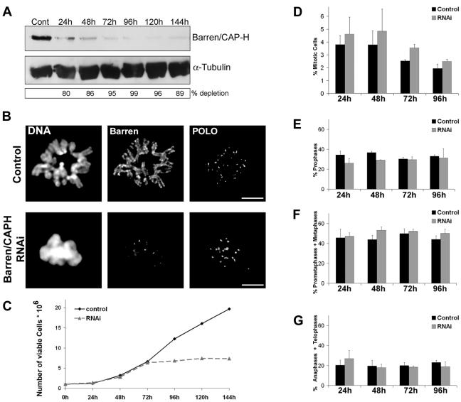
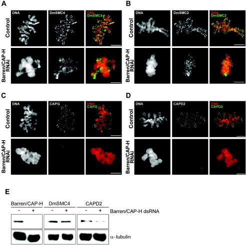
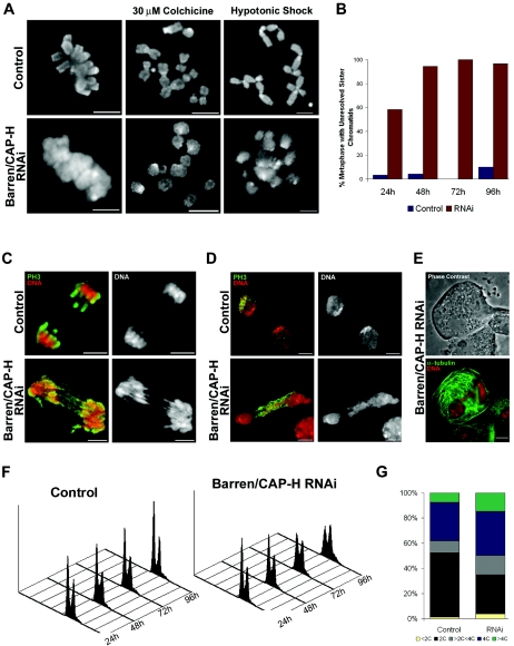
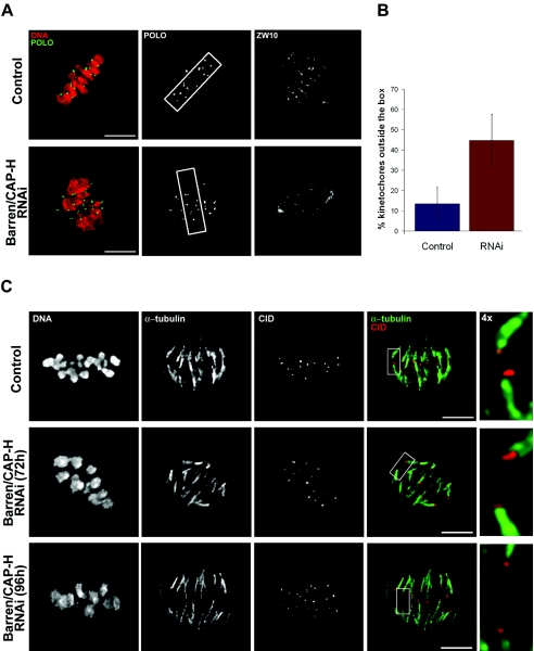
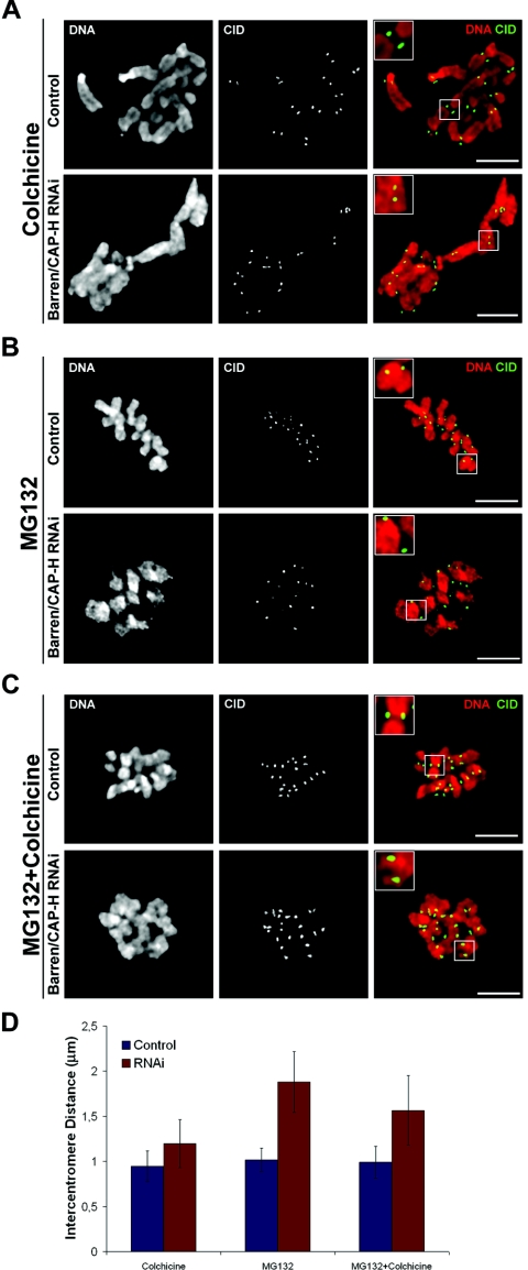
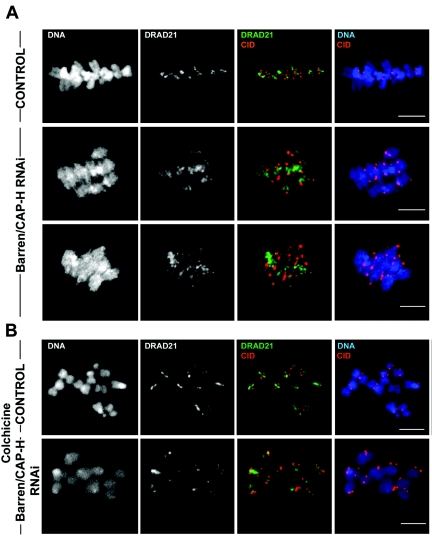
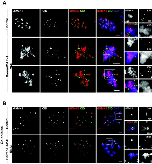
During cell division, chromatin undergoes structural changes essential to ensure faithful segregation of the genome. Condensins, abundant components of mitotic chromosomes, are known to form two different complexes, condensins I and II. To further examine the role of condensin I in chromosome structure and in particular in centromere organization, we depleted from S2 cells the Drosophila CAP-H homologue Barren, a subunit exclusively associated with condensin I. In the absence of Barren/CAP-H the condensin core subunits DmSMC4/2 still associate with chromatin, while the other condensin I non-structural maintenance of chromosomes family proteins do not. Immunofluorescence and in vivo analysis of Barren/CAP-H-depleted cells showed that mitotic chromosomes are able to condense but fail to resolve sister chromatids. Additionally, Barren/CAP-H-depleted cells show chromosome congression defects that do not appear to be due to abnormal kinetochore-microtubule interaction. Instead, the centromeric and pericentromeric heterochromatin of Barren/CAP-H-depleted chromosomes shows structural problems. After bipolar attachment, the centromeric heterochromatin organized in the absence of Barren/CAP-H cannot withstand the forces exerted by the mitotic spindle and undergoes irreversible distortion. Taken together, our data suggest that the condensin I complex is required not only to promote sister chromatid resolution but also to maintain the structural integrity of centromeric heterochromatin during mitosis.


Figures

FIG. 1.
FIG. 1.
Depletion of Barren/CAP-H from Schneider 2 cells by double-stranded RNA interference. (A) Total protein extracts from 5 × 10⁵ cells were run on a 7.5% SDS-PAGE and Barren/CAP-H protein levels were monitored at different times of the experiment by Western blot. α-Tubulin was used as loading control. (B) Depletion of Barren/CAP-H was confirmed by immunofluorescence. Metaphase chromosomes from double-stranded RNA-treated cells show no accumulation of Barren/CAP-H in contrast to control cells, where Barren/CAP-H is localized at the axis of each chromatid. Scale bars are 5 μm. (C) Proliferation profiles of control and Barren/CAP-H double-stranded RNA interference-treated S2 cells throughout the experiment. (D) Mitotic index and (F to H) mitotic progression throughout the experiment were quantified using either POLO/phospho-histone 3 or tubulin/phospho-histone 3 double staining. Approximately 6,500 cells were counted for each time point. Note that there is no significant change in both the mitotic index and mitotic progression between control and Barren/CAP-H-depleted cells meaning that Barren/CAP-H-depleted cells are able to undergo mitosis with normal timing.
FIG. 2.
FIG. 2.
Stability of condensin I after depletion of Barren/CAP-H. (A to D) Immunolocalization of condensin subunits in control and Barren/CAP-H-depleted (96 h) S2 cells. In control metaphase cells, all other condensin I subunits, including (A) DmSMC4, (B) DmSMC2, (C) DmCAP-D2, and (D) DmCAP-G, localize at the axis of sister chromatids. After depletion of Barren/CAP-H, DmSMC4 and DmSMC2 still localize to chromatin but now appear diffused and are no longer confined to the axis of chromosomes. However, the non-SMC subunits DmCAP-D2 and DmCAP-G are not able to localize to Barren/CAP-H-depleted chromosomes. Scale bars are 5 μm. (E) Total protein extracts from 106 cells were assayed by Western blot to determine the levels of DmSMC4 and CAP-D2 in control (−) or Barren/CAP-H RNAi-treated (+) cells. Note that the levels of DmSMC4 do not change significantly after Barren/CAP-H depletion compared to the controls. In contrast, DmCAP-D2 levels are significantly reduced (45%) compared to the controls. Tubulin was used as the loading control and quantifications were performed using Image J Software.
FIG. 3.
FIG. 3.
Characterization of S2 cells after depletion of Barren/CAP-H. (A) Control or Barren/CAP-H-depleted cells were fixed directly after 96 h or treated with colchicine (2 h) or hypotonic shock before fixation and then stained with DAPI to reveal chromosome structure. Unlike the chromosome morphology in control cells, Barren/CAP-H-depleted chromosomes show unresolved sister chromatids that become fuzzy after hypotonic shock. (B) Quantification of the percentage of cells in metaphase with unresolved sister chromatids shows that at 96 h virtually all cells in metaphase show unresolved sister chromatids. (C and D) Depletion of Barren/CAP-H leads to the formation of DNA bridges observed in anaphase and telophase. Cells were immunostained with an anti-phospho-histone H3 antibody (PH3) and DNA. (E) Representative image of a giant binucleated cell found after Barren/CAP-H depletion. Cells were immunostained for α-tubulin and DNA. Scale bars are 5 μm. (F) FACS profiles of both control and Barren/CAP-H-depleted cells, showing DNA content and cell number at different time points. (G) Graphic representation of the frequency of cells with different DNA content obtained from FACS analysis at 96 h after double-stranded RNA addition.
FIG. 4.
FIG. 4.
In vivo analysis of mitotic progression after depletion of Barren/CAP-H. Selected images from time-lapse movies of control and Barren/CAP-H-depleted (72 h) S2 cells stably expressing GFP-histone H2B acquired every 2 min from the time mitotic chromosomes could be identified. To align the movies anaphase onset was defined as time zero. (A) In control cells, prometaphase is followed by a tight organization of the chromosome at the metaphase plate, which after a few minutes initiates sister chromatid separation. (B) Analysis of Barren/CAP-H-depleted cells shows that these cells never appear to define a normal tight metaphase plate. Furthermore, chromatin bridges are observed as soon as anaphase is initiated. (C) In some cases we also observed cells depleted of Barren/CAP-H undergo anaphase onset but extensive chromatin bridges form, and after an initial attempt to segregate, the chromatin collapses back into a single large nucleus.
FIG. 5.
FIG. 5.
Kinetochore microtubule attachment and congression after depletion of Barren/CAP-H. (A) Both control and Barren/CAP-H-depleted cells (96 h) were arrested in metaphase by incubation with the proteasome inhibitor MG132 for 2 h. In order to evaluate kinetochore congression, a box comprising 85% of aligned kinetochores in a control metaphase was defined. The same box was placed over Barren/CAP-H-depleted metaphases perpendicular to the spindle (indicated by ZW10 spindle staining). (B) Quantification of the percentage of kinetochores placed outside the box in both control and Barren/CAP-H-depleted metaphase cells (for the control 14 cells were analyzed, n = 292 kinetochores, and for Barren/CAP-H depletion 14 cells were analyzed, n = 268 kinetochores). Note that depletion of Barren/CAP-H causes a severe increase in the frequency of misaligned kinetochores. (C) Control or Barren/CAP-H-depleted cells arrested with MG132 for 2 h were incubated with calcium to remove all microtubules except the kinetochore fiber. Cells were fixed and immunostained for α-tubulin and CID. Higher magnification images (4×) show that in both control and Barren/CAP-H-depleted cells, metaphase chromosomes are under bipolar attachment. Scale bars are 5 μm.
FIG. 6.
FIG. 6.
Analysis of intercentromere distances after depletion of Barren/CAP-H. (A to C) Both control and Barren/CAP-H-depleted (72 h) cells were immunostained for CID and DNA. Cultures were (A) incubated with 30 μM colchicine for 2 h to depolymerize all microtubules before entering mitosis, (B) incubated for 2 h with 20 μM MG132 to arrest cells in metaphase, (C) incubated with 20 μM MG132 to arrest cells in metaphase followed by a 30-min incubation with 30 μM colchicine to depolymerize all microtubules that were previously attached to the kinetochores. Scale bars are 5 μm. Higher magnifications (2×). (D) Quantification of intercentromere distances of control and Barren/CAP-H-depleted cells after the indicated experimental conditions.
FIG. 7.
FIG. 7.
Analysis of the localization of SCC1/DRAD21 on chromosomes after depletion of Barren/CAP-H. Both control and Barren/CAP-H-depleted (96 h) cells were immunostained for POLO and DRAD21. (A) Cells arrested at metaphase by 2 h incubation with 20 μM MG132. In control cells SCC1/DRAD21 localizes between sister chromatids as a tight line between sister centromeres. However, after depletion of Barren/CAP-H, SCC1/DRAD21 is distributed over a broad area between sister centromeres. (B) Cells were incubated with 30 μM colchicine for 2 h to arrest them at prometaphase before microtubules could bind kinetochores. In these cells, SCC1/DRAD21 localizes to a thin line between sister centromeres in both control and Barren/CAP-H-depleted chromosomes. Scale bars are 5 μm.
FIG. 8.
FIG. 8.
Immunolocalization of dimethylated K9 histone H3 in the heterochromatin of Barren/CAP-H RNAi-depleted chromosomes. Control and Barren/CAP-H cells were immunostained for CID and K9 dimethylated histone H3 (diMeK9). (A) In control cells at metaphase, diMeK9 is confined to heterochromatin. However, after depletion of Barren/CAP-H the pattern of diMeK9 appears to be significantly altered under bipolar attachment. diMeK9 is now detected over a broad area of chromatin localized between the two CID-labeled centromeres. (B) Both control and Barren-CAP-H-depleted cells were incubated with 30 μM colchicine for 2 h to depolymerize microtubules and the pattern of diMeK9 was determined. Note that under these conditions diMeK9 is confined to a tight region between sister centromeres in both control and Barren/CAP-H-depleted cells. Scale bars are 5 μm.

Similar articles

Cited by

References

    1. Adams, R. R., H. Maiato, W. C. Earnshaw, and M. Carmena. 2001. Essential roles of Drosophila inner centromere protein (INCENP) and aurora B in histone H3 phosphorylation, metaphase chromosome alignment, kinetochore disjunction, and chromosome segregation. J. Cell Biol. 153:865-880. - PMC - PubMed
    1. Almagro, S., D. Riveline, T. Hirano, B. Houchmandzadeh, and S. Dimitrov. 2004. The mitotic chromosome is an assembly of rigid elastic axes organized by structural maintenance of chromosomes (SMC) proteins and surrounded by a soft chromatin envelope. J. Biol. Chem. 279:5118-5126. - PubMed
    1. Aono, N., T. Sutani, T. Tomonaga, S. Mochida, and M. Yanagida. 2002. Cnd2 has dual roles in mitotic condensation and interphase. Nature 417:197-202. - PubMed
    1. Bhat, M. A., A. V. Philp, D. M. Glover, and H. J. Bellen. 1996. Chromatid segregation at anaphase requires the barren product, a novel chromosome-associated protein that interacts with topoisomerase II. Cell 87:1103-1114. - PubMed
    1. Bickmore, W. A., and K. Oghene. 1996. Visualizing the spatial relationships between defined DNA sequences and the axial region of extracted metaphase chromosomes. Cell 84:95-104. - PubMed

Publication types

MeSH terms

LinkOut - more resources