. 2005 Oct;25(20):9016-27.
doi: 10.1128/MCB.25.20.9016-9027.2005.

Regulating SWI/SNF subunit levels via protein-protein interactions and proteasomal degradation: BAF155 and BAF170 limit expression of BAF57

Affiliations

Regulating SWI/SNF subunit levels via protein-protein interactions and proteasomal degradation: BAF155 and BAF170 limit expression of BAF57

Jianguang Chen et al. Mol Cell Biol. 2005 Oct.

Abstract

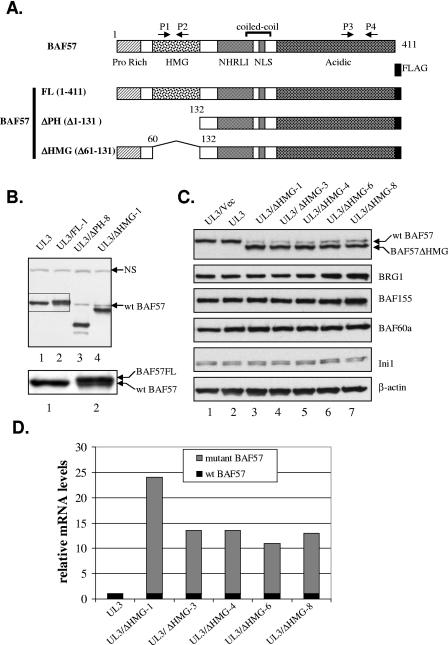
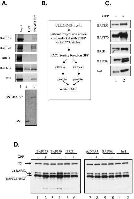
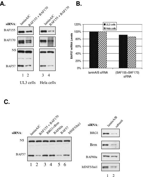
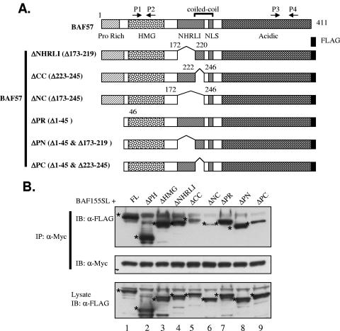
The mammalian SWI/SNF chromatin remodeling complex, whose function is of critical importance in transcriptional regulation, contains approximately 10 protein components. The expression levels of the core SWI/SNF subunits, including BRG1/Brm, BAF155, BAF170, BAF60, hSNF/Ini1, and BAF57, are stoichiometric, with few to no unbound molecules in the cell. Here we report that exogenous expression of the wild type or certain deletion mutants of BAF57, a key subunit that mediates the interaction between the remodeling complex and transcription factors, results in diminished expression of endogenous BAF57. This down-regulation process is mediated by an increase in proteasome-dependent degradation of the BAF57 protein. Furthermore, the protein levels of BAF155/170 dictate the maximum cellular amount of BAF57. We mapped the domains responsible for the interaction between BAF57 and BAF155 and demonstrated that protein-protein interactions between them play an important role in this regulatory process. These findings provide insights into the physiological mechanisms responsible for maintaining the proper stoichiometric levels of the protein components comprising multimeric enzyme complexes.


Figures

FIG. 1.
FIG. 1.
Exogenous expression of BAF57 down-regulates endogenous BAF57. (A) Schematic representation of human BAF57 domain structure and the FLAG-tagged constructs used in this study. Arrows mark the positions of real-time PCR primer pairs. HMG, high mobility group; NLS, nuclear localization signal. (B) Whole-cell extracts (WCEs) from parental UL3 cells (lane 1) and UL3-derived cell lines expressing either full-length BAF57 (lane 2) or the BAF57ΔPH (lane 3) or BAF57ΔHMG (lane 4) mutant were analyzed by Western blotting with anti-BAF57 antibody. The boxed area was enlarged and is shown under the main figure to illustrate the separation of wt BAF57 and BAF57FL proteins. “NS” denotes a nonspecific band recognized by the BAF57 antibody and indicates equivalent loading. (C) WCEs from parental UL3 cells (lane 2) and UL3-derived cell lines expressing either an empty vector (lane 1) or the BAF57ΔHMG mutant (lanes 3 to 7) were analyzed by Western blotting for the expression of BAF57, other SWI/SNF subunits, and β-actin. (D) Relative mRNA levels of wt BAF57 and BAF57ΔHMG in UL3 cells and various UL3/BAF57ΔHMG cell lines, as determined by quantitative RT-PCR. The relative wt BAF57 mRNA level was determined with the P1/P2 primer set. The relative amount of wt BAF57 mRNA in UL3 cells as well as the amount of combined wt and ΔHMG BAF57 mRNAs in various UL3/BAF57ΔHMG cell lines was first determined with the primer set P3/P4 and then adjusted according to the amplification efficiency difference between P1/P2 and P3/P4, as estimated by comparing CT values of P1/P2- and P3/P4-directed amplification of equal amounts of mRNA from UL3 cells. An arbitrary value of 1 was assigned to the wt BAF57 mRNA level in UL3 cells determined with the P1/P2 primers. All other values for a given cell line are presented relative to this value in the graph.
FIG. 2.
FIG. 2.
Inhibition of proteasome and translation has distinct effects on the BAF57 protein level in UL3 and UL3/BAF57ΔHMG-1 cells. UL3/BAF57ΔHMG-1 cells were treated with either MG132 (A), epoxomycin (B), or cycloheximide (C) for 0, 2, 4, 6, and 8 h. Whole-cell extracts were prepared at each time point and resolved by 12% SDS-PAGE. The relative protein levels of wt BAF57 and BAF57ΔHMG in the extracts were determined by Western blotting. UL3 cells were also treated with either MG132 (D) or cycloheximide (E) for 0, 2, 4, 6, and 8 h. The relative protein level of wt BAF57 present at each time point was again determined by Western blotting. “NS” denotes a nonspecific band recognized by the BAF57 antibody and indicates equivalent loading.
FIG. 3.
FIG. 3.
BAF155/170 interacts with BAF57 and increases steady-state levels of BAF57 protein in UL3/BAF57ΔHMG-1 cells. (A) Pull-down assay of core SWI/SNF subunits translated in vitro by GST-BAF57. The 35S-labeled subunit proteins were incubated with either GST (lane 2) or GST-BAF57 (lane 3), and bound proteins were detected with a phosphorimager (upper panel). A Coomassie-stained gel of GST and the GST-BAF57 fusion protein is shown in the lower panel. (B) Strategy for enriching UL3/BAF57ΔHMG-1 cells transfected with pcDNA3 or plasmids expressing various core SWI/SNF subunits. (C) WCEs from GFP-negative (lane 1) and -positive (lane 2) cells after transfection were analyzed by Western blotting with antibodies to core SWI/SNF subunits. (D) The same extracts used in panel C as well as WCEs from pcDNA3-transfected cells were analyzed by Western blotting with BAF57 antibody. “NS” denotes a nonspecific band and indicates equivalent loading.
FIG. 4.
FIG. 4.
Reduction of BAF155 and BAF170 expression decreases BAF57 protein level in UL3 and HeLa cells. (A) UL3 and HeLa cells were transfected with lamin A/C siRNA (lanes 1 and 3) or a mixture of BAF155 and BAF170 siRNAs (lanes 2 and 4). Whole-cell extracts were prepared 48 h after transfection and analyzed by Western blotting for the expression of BAF155, BAF170, and BAF57. (B) UL3 and HeLa cells were treated as described for panel A, but total RNA was prepared from those cells, and the relative mRNA level of wt BAF57 was determined with the P1/P2 primer set by real-time PCR. The BAF57 mRNA level in UL3 and HeLa cells transfected with the control siRNA was arbitrarily set at 100%. (C) HeLa cells were transfected with either lamin siRNA (lane 1) or siRNAs targeting BAF155/170, BRG1/Brm, BAF60a, BAF57, and hSNF5/Ini1, respectively (lanes 2 to 6). Whole-cell extracts were prepared 48 h after transfection and analyzed by Western blotting for the expression of BAF57 (left panel). The same extracts were also probed with antibodies to BRG1/Brm, BAF60a, and hSNF5/Ini1 to show the knockdown efficiency (right panel).
FIG. 5.
FIG. 5.
Specific BAF155 domains are required for increasing the BAF57 protein level and the interaction with BAF57 in vivo. (A) Schematic illustration of human BAF155 domain structure and the various deletion constructs used in this study. All mutants were fused to a nuclear localization signal (NLS) and Myc epitope at the C terminus. P/Q, proline- and glutamine-rich region; LZ, leucine zipper. (B) Whole-cell extracts from UL3/BAF57ΔHMG-1 cells transiently transfected with either an empty vector (lane a), wt BAF155 (lane b), or various deletion mutant expression vectors (lanes c to i) were analyzed by Western blotting with either BAF57 antibody (top panel) or Myc antibody (bottom panel). β-Actin was used as an internal loading control (middle panel). (C) Whole-cell extracts were prepared from UL3 cells transiently transfected with BAF57-FLAG and various BAF155 mutant expression vectors and immunoprecipitated with Myc antibody. The precipitated materials were analyzed by Western blotting with FLAG-horseradish peroxidase (FLAG-HRP) antibody (top panel). The same blot was stripped and reprobed with Myc-HRP antibody (middle panel) to demonstrate the IP efficiency. The bands corresponding to the expected BAF155 protein are marked with dots. About 5% of the total input lysates used for immunoprecipitation were directly analyzed by Western blotting with FLAG antibody (bottom panel).
FIG. 6.
FIG. 6.
Conserved BAF57 domains are required for interaction with BAF155 in vivo. (A) Schematic description of BAF57 deletion mutants used in this study. (B) Whole-cell extracts were prepared from UL3 cells transiently transfected with the BAF155SL expression plasmid (Fig. 5A) and various BAF57 deletion constructs and then were immunoprecipitated with Myc antibody. The precipitated materials were analyzed by Western blotting with FLAG-HRP antibody (top panel). The same blot was stripped and reprobed with Myc-HRP antibody (middle panel) to demonstrate the IP efficiency. About 5% of the total input lysates used for immunoprecipitation were directly analyzed by Western blotting with FLAG antibody (bottom panel). The band corresponding to the expected protein in each lane is marked with an asterisk.

References

    1. Aasland, R., A. F. Stewart, and T. Gibson. 1996. The SANT domain: a putative DNA-binding domain in the SWI-SNF and ADA complexes, the transcriptional co-repressor N-CoR and TFIIIB. Trends Biochem. Sci. 21:87-88. - PubMed
    1. Aravind, L., and L. M. Iyer. 24July2002, posting date. The SWIRM domain: a conserved module found in chromosomal proteins points to novel chromatin-modifying activities. Genome Biol. 3:RESEARCH 0039. [Online.] http://genomebiology.com/2002/3/8/research0039. - PMC - PubMed
    1. Baker, K. M., G. Wei, A. E. Schaffner, and M. C. Ostrowski. 2003. Ets-2 and components of mammalian SWI/SNF form a repressor complex that negatively regulates the BRCA1 promoter. J. Biol. Chem. 278:17876-17884. - PubMed
    1. Belandia, B., R. L. Orford, H. C. Hurst, and M. G. Parker. 2002. Targeting of SWI/SNF chromatin remodelling complexes to estrogen-responsive genes. EMBO J. 21:4094-4103. - PMC - PubMed
    1. Boyer, L. A., R. R. Latek, and C. L. Peterson. 2004. The SANT domain: a unique histone-tail-binding module? Nat. Rev. Mol. Cell Biol. 5:158-163. - PubMed

Publication types

MeSH terms