. 2005 Oct;25(20):9028-39.
doi: 10.1128/MCB.25.20.9028-9039.2005.

The Xenopus ELAV protein ElrB represses Vg1 mRNA translation during oogenesis

Affiliations

The Xenopus ELAV protein ElrB represses Vg1 mRNA translation during oogenesis

Lucy J Colegrove-Otero et al. Mol Cell Biol. 2005 Oct.

Abstract

Xenopus laevis Vg1 mRNA undergoes both localization and translational control during oogenesis. We previously characterized a 250-nucleotide AU-rich element, the Vg1 translation element (VTE), in the 3'-untranslated region (UTR) of this mRNA that is responsible for the translational repression. UV-cross-linking and immunoprecipitation experiments, described here, revealed that the known AU-rich element binding proteins, ElrA and ElrB, and TIA-1 and TIAR interact with the VTE. The levels of these proteins during oogenesis are most consistent with a possible role for ElrB in the translational control of Vg1 mRNA, and ElrB, in contrast to TIA-1 and TIAR, is present in large RNP complexes. Immunodepletion of TIA-1 and TIAR from Xenopus translation extract confirmed that these proteins are not involved in the translational repression. Mutagenesis of a potential ElrB binding site destroyed the ability of the VTE to bind ElrB and also abolished translational repression. Moreover, multiple copies of the consensus motif both bind ElrB and support translational control. Therefore, there is a direct correlation between ElrB binding and translational repression by the Vg1 3'-UTR. In agreement with the reporter data, injection of a monoclonal antibody against ElrB into Xenopus oocytes resulted in the production of Vg1 protein, arguing for a role for the ELAV proteins in the translational repression of Vg1 mRNA during early oogenesis.


Figures

FIG. 1.
FIG. 1.
Depiction of the constructs utilized. The inserts shown were cloned into the SacI and BamHI sites of the firefly luciferase vector (pLUC) and pGEM1 to give the appropriate constructs. The −1200 insert consisted of a 1.2-kb fragment derived from the vector pSP64. The other inserts shown were derived from the Vg1 3′-UTR, and their relative positions within the 3′-UTR are depicted. The asterisk shows the position of the ElrB consensus binding motif that is mutated in the VUmut and VTEmut constructs.
FIG. 2.
FIG. 2.
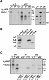
ElrA and ElrB and TIA-1 and TIAR interact with the VTE. (A) The left panel shows immunoprecipitation, performed using an antibody against HuR, of UV-cross-linking reactions containing 32P-labeled VTE probe or VLE probe as a control. The proteins were detected by autoradiography. The three lanes for each RNA show the original UV-cross-linking reaction (UV), the immunoprecipitated sample (IP), and the proteins remaining in the supernatant (SN). The proteins FRGY2 and Vg1RBP, previously identified by immunoprecipitation (42), are shown in the UV sample. Molecular size markers are shown in kilodaltons. The right panel shows a Western blot of the VTE samples with anti-HuR. (B) Western analysis with anti-HuR (α-HuR) of immunoprecipitations from stage II lysate, performed using antipeptide antibodies directed specifically against ElrB (α-ElrB) or ElrA (α-ElrA). Lanes labeled α-ElrB PI and α-ElrA PI show immunoprecipitations performed using the relevant preimmune sera. IgG, immunoglobulin G. (C) Immunoprecipitation of UV-cross-linking reactions with labeled VTE and VLE, performed using antibodies directed against TIA-1 (α-TIA-1) and TIAR (α-TIAR). The immunoprecipitated proteins (IP) and proteins remaining in the supernatant (SN) are shown in each case. Molecular size markers are shown in kilodaltons.
FIG. 3.
FIG. 3.
Characterization of ElrA, ElrB, TIA-1, and TIAR from Xenopus oocytes. (A) Western analysis of lysates prepared with oocytes from the six stages of oogenesis (I through VI shown at top), performed using anti-HuR (α-HuR), anti-TIAR (α-TIAR), and anti-Vg1 (α-Vg1) antibodies and anti-Xp54 (α-Xp54) antibody as a control. (B) Western analysis of nuclear (N) and cytoplasmic (C) extracts, performed using anti-HuR and anti-TIAR, with anti-PARN (α-PARN) as a control for the integrity of the nuclear and cytoplasmic fractions. (C) Western analysis of fractions of stage III extract obtained from a Superose 6 HR 10/30 gel filtration column. Gel filtration was also performed with extract which had been pretreated with RNase A. Size estimations were obtained from a calibration curve obtained using thyroglobulin (669 kDa), ferritin (440 kDa), catalase (232 kDa), aldolase (158 kDa), ovalbumin (43 kDa), and chymotrypsinogen A (25 kDa). Lanes T show samples of nonfiltrated extract.
FIG. 4.
FIG. 4.
Immunodepletion of Xenopus translation extract. (A) Western analysis of mock-depleted and anti-HuR-depleted translation extract, using anti-HuR as the primary antibody. (B) Western analysis with anti-TIAR of mock-depleted and anti-TIAR-depleted translation extract. (C) Three different luciferase reporter RNAs (LUC-1200, LUC-VU, and LUC-VU1) were assayed in the mock-depleted and anti-TIAR-depleted translation extracts. (D) RT-PCR, of total RNA extracted from immunoprecipitations from stage II oocytes carried out using the indicated antibodies, was performed with primers specific to the Vg1 3′-UTR. The expected size of the fragment is 420 bp. Lanes M contain molecular size markers. Abbreviations: SN, proteins remaining in the supernatant; Prot. A-Seph., protein A-Sepharose alone; Prot. A-Seph. + α-HuR, protein A-Sepharose beads preincubated with anti-HuR antibody; Prot. G-Seph., protein G-Sepharose alone; Prot. G-Seph. + α-TIAR, protein G-Sepharose beads preincubated with anti-TIAR antibody; IgG, immunoglobulin G; Ab, antibody; PI, immunoprecipitations using the relevant preimmune sera; Temp., template; RT, reverse transcriptase; Fluc, firefly luciferase; Rluc, Renilla luciferase.
FIG. 5.
FIG. 5.
Mutation of a putative ElrB binding site markedly reduces ElrB binding to the VTE. (A) An alignment of the 5′ AU-rich regions from X. tropicalis (T; EST TEgg016d06 5′ [http://www.sanger.ac.uk/cgi-bin/BLAST/submitblast/x_tropicalis]), X. borealis (B; AFO41844), and X. laevis (L; M18055) is shown. Conserved nucleotides are underlined in the X. laevis sequence. A conserved motif highlighted in bold corresponds to the ElrB homologue binding site described by Gao et al. (17). This motif is mutated to the italicized sequence in the VUmut and VTEmut constructs. (B) UV-cross-linking reactions performed with either 32P-labeled VTE or VTEmut are shown. Lane M, molecular size markers are shown in kilodaltons. (C) Band shift assays were performed using 32P-labeled VTE or VTEmut and total ovary extract. Anti-HuR (α-HuR) antibody was added to determine whether the shifted complexes contained ElrA or ElrB.
FIG. 6.
FIG. 6.
Mutation of the ElrB binding site abolishes translational repression by the VTE. (A) Luciferase reporter mRNAs were injected into stage VI oocytes, and the resulting luciferase activities were assayed. (B) The same reporter RNAs were analyzed with Xenopus translation extract. Both panels A and B show results representative of at least three independent experiments. (C) The stability of the reporter RNAs was measured in extract. The top panel shows a comparison of 32P-labeled LUC-1200, LUC-VU, and LUC-mut RNAs extracted from translation extract at the given times and separated by agarose gel electrophoresis. The bottom panel shows ethidium bromide staining of rRNA as a loading control. Fluc, firefly luciferase; Rluc, Renilla luciferase.
FIG. 7.
FIG. 7.
Multiple copies of the ElrB binding site enable ElrB binding and translational repression. (A) UV-cross-linking reactions were performed with 32P-labeled VTE, VU1, VTEmut, and 5×EBS RNAs (UV). The right side of the panel shows immunoprecipitation of these UV-cross-linking reactions, performed using anti-HuR antibody (IP). Molecular size markers are given in kilodaltons. (B) Luciferase reporter RNAs were injected into stage VI oocytes, and the resulting luciferase activities were assayed. (C) Luciferase reporter RNAs were assayed in Xenopus translation extract. Both panels B and C show results representative of at least three independent experiments. (D) The stability of the reporter RNAs was measured in extract. The top panel shows a comparison of 32P-labeled LUC-1200 and LUC-5×EBS RNAs extracted from translation extract at the given times and separated by agarose gel electrophoresis. The bottom panel shows ethidium bromide staining of rRNA as a loading control. No degradation was observed during the time course of the experiment. (E) Xenopus translation extract was preincubated with increasing amounts of 5×EBS competitor RNA. The preincubated aliquots of extract were then utilized for translation assays with the reporter RNAs shown, and the resulting luciferase activities were measured. The amounts of 5×EBS RNA added were in molar excess, as shown, over the reporter RNAs. Fluc, firefly luciferase; Rluc, Renilla luciferase.
FIG. 8.
FIG. 8.
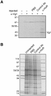
Injection of anti-HuR antibody alleviates Vg1 translational repression. (A) Immunoprecipitation of [35S]methionine-labeled stage III oocytes using anti-Vg1 (α-Vg1) antibody. Prior to labeling, the oocytes were injected with either phosphate-buffered saline (PBS) buffer or an equal amount of either a control antibody (Ab) or anti-HuR (α-HuR) antibody. Molecular size markers are shown in kilodaltons on the left. (B) Lysates prepared from 35S-labeled oocytes were subjected to SDS-PAGE and autoradiography. Each lane represents one oocyte equivalent. This experiment was performed twice, with similar results.

Similar articles

Cited by

References

    1. Antic, D., and J. D. Keene. 1997. Embryonic lethal abnormal visual RNA-binding proteins involved in growth, differentiation and posttranscriptional gene expression. Am. J. Hum. Genet. 61:273-278. - PMC - PubMed
    1. Antic, D., N. Lu, and J. D. Keene. 1999. ELAV tumor antigen, Hel-N1, increases translation of neurofilament M mRNA and induces formation of neurites in human teratocarcinoma cells. Genes Dev. 13:449-461. - PMC - PubMed
    1. Atasoy, U., J. Watson, D. Patel, and J. D. Keene. 1998. ELAV protein HuA (HuR) can redistribute between nucleus and cytoplasm and is upregulated during serum stimulation and T cell activation. J. Cell Sci. 111:3145-3156. - PubMed
    1. Campos, A. R., D. Grossman, and K. White. 1985. Mutant alleles at the locus elav in Drosophila melanogaster lead to nervous system defects. A developmental-genetic analysis. J. Neurogenet. 2:197-218. - PubMed
    1. Chen, C.-Y., and A.-B. Shyu. 1995. AU-rich elements: characterisation and importance in mRNA degradation. Trends Biochem. Sci. 20:465-470. - PubMed

Publication types

MeSH terms