. 2005 Oct;115(10):2762-73.
doi: 10.1172/JCI24381.

Deficits in amygdaloid cAMP-responsive element-binding protein signaling play a role in genetic predisposition to anxiety and alcoholism

Affiliations

Deficits in amygdaloid cAMP-responsive element-binding protein signaling play a role in genetic predisposition to anxiety and alcoholism

Subhash C Pandey et al. J Clin Invest. 2005 Oct.

Erratum in

  • J Clin Invest. 2006 Dec;116(12):3292

Abstract

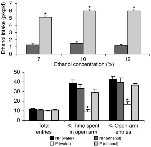
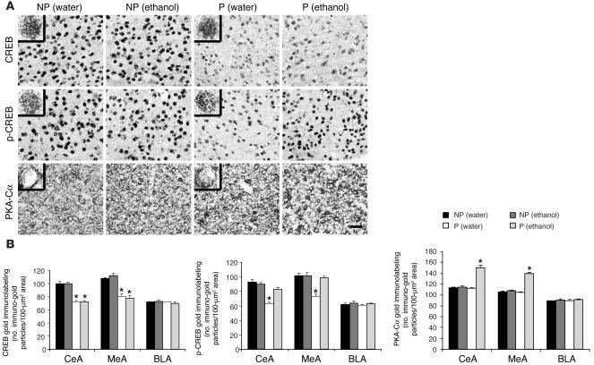
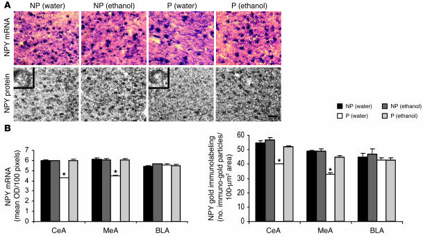
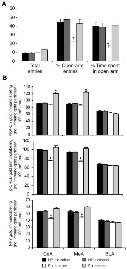
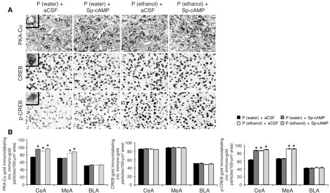
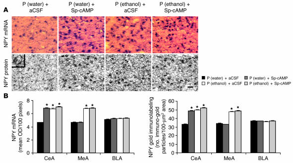
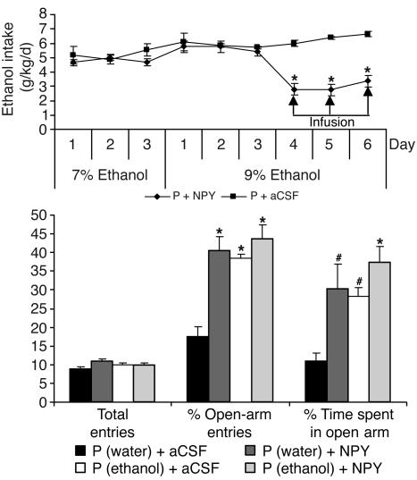
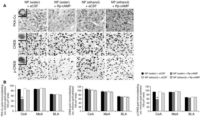
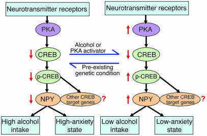
We investigated the role of cAMP-responsive element-binding protein (CREB) in genetic predisposition to anxiety and alcohol-drinking behaviors using alcohol-preferring (P) and -nonpreferring (NP) rats. The levels of CREB, phosphorylated CREB, and neuropeptide Y (NPY) were innately lower in the central amygdala (CeA) and medial amygdala (MeA), but not in the basolateral amygdala (BLA), of P rats compared with NP rats. P rats displayed higher baseline anxiety-like behaviors and consumed higher amounts of alcohol compared with NP rats. Ethanol injection or voluntary intake reduced the higher anxiety levels in P rats. Ethanol also increased CREB function in the CeA and MeA, but not in the BLA, of P rats. Infusion of the PKA activator Sp-cAMP or NPY into the CeA decreased the alcohol intake and anxiety-like behaviors of P rats. PKA activator infusion also increased CREB function in the CeA of P rats. On the other hand, ethanol injection or voluntary intake did not produce any changes either in anxiety levels or on CREB function in the amygdaloid structures of NP rats. Interestingly, infusion of the PKA inhibitor Rp-cAMP into the CeA provoked anxiety-like behaviors and increased alcohol intake in NP rats. PKA inhibitor decreased CREB function in the CeA of NP rats. These novel results provide the first evidence to our knowledge that decreased CREB function in the CeA may be operative in maintaining the high anxiety and excessive alcohol-drinking behaviors of P rats.


Figures

Figure 1
Figure 1
Alcohol-drinking patterns and anxiety-like behaviors in P and NP rats. Upper panel: Ethanol intake (7% for 3 days, 10% for 3 days, and 12% for 3 days) was measured by the 2-bottle free choice paradigm in P and NP rats. Values represent the mean ± SEM of 6 rats in each group. Lower panel: The effects of voluntary ethanol intake on open- and closed-arm activities of P and NP rats in the EPM test. Values represent the mean ± SEM of 5–6 rats in each group. *P < 0.001 versus respective controls.
Figure 2
Figure 2
Voluntary ethanol intake and CREB signaling in the amygdala of P and NP rats. (A) Low-magnification views of CREB, p-CREB, and PKA-Cα gold immunolabeling in CeA structures of P and NP rats with or without ethanol exposure. Photomicrographs show CREB-positive nuclei (upper panels), p-CREB–positive nuclei (middle panels), and PKA-Cα–positive cell bodies (lower panels) in the CeA of NP and P rats with or without ethanol exposure. Insets show the immuno-gold particles within a single nucleus (CREB and p-CREB) or cell body (PKA-Cα) at high magnification (×100). Scale bar: 40 μm. (B) Effect of voluntary ethanol intake on CREB, p-CREB, and PKA-Cα protein levels in various amygdaloid structures (CeA, MeA, and BLA) of P and NP rats. Values represent the mean ± SEM of 4–6 rats in each group. *P < 0.001 versus respective controls.
Figure 3
Figure 3
Voluntary ethanol intake and NPY levels in the amygdala of P and NP rats. (A) Low-magnification views of NPY mRNA expression (in situ RT-PCR; upper panels) and NPY gold immunolabeling (lower panels) in the CeA of NP and P rats with or without ethanol exposure. Insets show the immuno-gold particles in a cell body at high magnification (×100). Scale bar: 40 μm. (B) Effects of voluntary ethanol intake on mRNA and protein levels of NPY in the CeA, MeA, and BLA structures of NP and P rats. Values represent the mean ± SEM of 4–6 rats in each group. *P < 0.001 versus respective controls.
Figure 4
Figure 4
Anxiety-like behaviors and CREB function in the amygdala of P and NP rats following acute ethanol exposure. (A) The effects of acute ethanol injection (i.p.) on open- and closed-arm activities of P and NP rats in the EPM test. Values represent the mean ± SEM of 5–7 rats in each group. (B) Effect of acute ethanol exposure on p-CREB, PKA-Cα, and NPY protein levels in various amygdaloid structures (CeA, MeA, and BLA) of P and NP rats. Values represent the mean ± SEM of 5 rats in each group. *P < 0.001 versus respective controls.
Figure 5
Figure 5
Low-magnification views of Nissl staining of brain sections showing the positions of cannula tips, probe lengths, and CeA target regions of P rats infused with aCSF or Sp-cAMP. Scale bar: 250 μm.
Figure 6
Figure 6
The effect of PKA activator infusion into the CeA on ethanol intake and anxiety-like behaviors in P rats. Upper panel: The alcohol-drinking patterns (7 and 9% ethanol) and the effect of Sp-cAMP infusion into the CeA on alcohol intake (last 3 days of 9% ethanol intake) in P rats. Values represent the mean ± SEM of 5–6 rats in each group. Lower panel: The effect of PKA activator infusion (0.5 μl of 80 nmol of Sp-cAMP or aCSF once daily for 3 days) and ethanol exposure on open- and closed-arm activities of P rats in the EPM. Values represent the mean ± SEM of 5–6 rats in each group. #P < 0.01, *P < 0.001 versus P rats infused with aCSF.
Figure 7
Figure 7
PKA activator infusion into the CeA during ethanol intake and CREB signaling in the amygdala of P rats. (A) Low-magnification views of PKA-Cα, CREB, and p-CREB gold immunolabeling in the CeA of P rats infused with or without Sp-cAMP during ethanol exposure. Photomicrographs show PKA-Cα–positive cell bodies or CREB- and p-CREB–positive nuclei in the CeA of P rats infused with aCSF or PKA activator. Insets show the immuno-gold particles within a single nucleus (CREB and p-CREB) or cell body (PKA-Cα) at high magnification (×100). Scale bar: 40 μm. (B) Effect of Sp-cAMP infusion into the CeA and voluntary ethanol exposure on PKA-Cα, CREB, and p-CREB protein levels in amygdaloid structures of P rats. Values represent the mean ± SEM of 5–6 rats in each group. *P < 0.001 versus P rats infused with aCSF.
Figure 8
Figure 8
PKA activator infusion into the CeA during ethanol intake and NPY levels in the amygdala of P rats. (A) Low-magnification views of NPY mRNA expression (in situ RT-PCR; upper panels) and NPY gold immunolabeling (lower panels) in the CeA of P rats with or without PKA activator infusion during ethanol exposure. Inset shows the gold particles in a cell body at high magnification (×100). Scale bar: 40 μm. (B) Effects of voluntary ethanol exposure and PKA activator infusion into the CeA on mRNA and protein levels of NPY in the CeA, MeA, and BLA structures of rats. Values represent the mean ± SEM of 5–6 rats in each group. *P < 0.001 versus respective controls.
Figure 9
Figure 9
The effect of NPY infusion into the CeA on ethanol intake and anxiety-like behaviors in P rats. Upper panel: The alcohol-drinking patterns (7 and 9% ethanol) and the effect of NPY infusion into the CeA (last 3 days of 9% ethanol intake) on alcohol intake in P rats. Values represent the mean ± SEM of 7 rats in each group. Lower panel: The effect of NPY infusion (0.5 μl of aCSF or 0.5 μl of 100 pmol of NPY, once daily for 3 days) and alcohol consumption on open- and closed-arm activities of P rats in the EPM. Values represent the mean ± SEM of 7 rats in each group. #P < 0.01, *P < 0.001 versus P rats infused with aCSF.
Figure 10
Figure 10
The effect of PKA inhibitor infusion into the CeA on ethanol intake and anxiety-like behaviors in NP rats. Upper panel: The alcohol-drinking patterns (7 and 9% ethanol) and the effect of Rp-cAMP infusion into the CeA (last 3 days of 9% ethanol intake) on alcohol intake in NP rats. Values represent the mean ± SEM of 7–8 rats in each group. Lower panel: The effect of PKA inhibitor infusion (0.5 μl of 40 nmol of Rp-cAMP or aCSF once daily for 3 days) and alcohol drinking on open- and closed-arm activities of NP rats in the EPM. Values represent the mean ± SEM of 7–8 rats in each group. #P < 0.01, *P < 0.001 versus NP rats infused with aCSF.
Figure 11
Figure 11
PKA inhibitor infusion into the CeA during ethanol intake and CREB signaling in the amygdala of NP rats. (A) Low-magnification views of PKA-Cα, CREB, and p-CREB gold immunolabeling in the CeA structures of NP rats infused with or without Rp-cAMP during ethanol exposure. Photomicrographs show PKA-Cα–positive cell bodies or CREB- and p-CREB–positive nuclei in the CeA of NP rats infused with aCSF or PKA inhibitor. Insets show the immuno-gold particles within a single nucleus (CREB and p-CREB) or cell body (PKA-Cα) at high magnification (×100). Scale bar: 40 μm. (B) Effect of Rp-cAMP infusion into the CeA and alcohol drinking on PKA-Cα, CREB, and p-CREB protein levels in amygdaloid structures of NP rats. Values represent the mean ± SEM of 5 rats in each group. *P < 0.001 versus NP rats infused with aCSF.
Figure 12
Figure 12
PKA inhibitor infusion into the CeA during ethanol intake and NPY levels in the amygdala of NP rats. (A) Low-magnification views of NPY mRNA expression (in situ RT-PCR; upper panels) and NPY gold immunolabeling (lower panels) in the CeA of NP rats with or without PKA inhibitor infusion during ethanol exposure. Inset shows the immuno-gold particles in a cell body at high magnification (×100). Scale bar: 40 μm. (B) Effects of voluntary ethanol exposure and PKA inhibitor infusion into the CeA on mRNA and protein levels of NPY in the CeA, MeA, and BLA structures of NP rats. Values represent the mean ± SEM of 5 rats in each group. *P <0.001 versus respective controls.
Figure 13
Figure 13
Molecular mechanisms in the neural circuitry of the CeA of P rats for genetic predisposition to anxiety and alcohol-drinking behaviors. The data collected in the present study suggest that decreased function of CREB (within the CREB→p-CREB→NPY system) in CeA neurocircuitry may be responsible for high anxiety levels and excessive alcohol-drinking behaviors of P rats. Thus, decreased CREB, p-CREB, and NPY levels represent a pre-existing genetic condition for anxiety and alcoholism. The ethanol exposure (blue arrows) causes an increase in active PKA and CREB phosphorylation and thereby increases NPY expression and attenuates anxiety behavior. PKA activator has the ability to mimic the action of alcohol on PKA and CREB phosphorylation and NPY expression in the CeA of P rats. NPY infusion into the CeA also attenuates anxiety and alcohol intake in P rats. CREB may also alter the expression of other CREB target genes (23), whose function needs to be evaluated in future studies of anxiety and alcohol-drinking behaviors in P rats. Red arrows indicate the increase or decrease of signaling protein levels.

Comment in

Similar articles

Cited by

References

    1. National Institute on Alcohol Abuse and Alcoholism. 1993. Eighth special report to the congress on alcohol and health. US Department of Health and Human Services, NIH. Bethesda, Maryland, USA.
    1. Enoch MA. Pharmacogenomics of alcohol response and addiction. Am. J. Pharmacogenomics. 2003;3:217–232. - PubMed
    1. Radel M, Goldman D. Pharmacogenetics of alcohol response and alcoholism: the interplay of genes and environmental factors in thresholds for alcoholism. Drug Metab. Dispos. 2001;29:489–494. - PubMed
    1. Thome J, Gewirtz JC, Weijers HG, Wiesbeck GA, Henn FA. Genome polymorphism and alcoholism. Pharmacogenomics. 2000;1:63–71. - PubMed
    1. Cloninger CR. Neurogenetic adaptive mechanisms in alcoholism. Science. 1987;236:410–416. - PubMed

Publication types

MeSH terms

Substances