Case Reports
. 2005 Oct;115(10):2784-92.
doi: 10.1172/JCI26020.

A molecular chaperone for mitochondrial complex I assembly is mutated in a progressive encephalopathy

Affiliations
Case Reports

A molecular chaperone for mitochondrial complex I assembly is mutated in a progressive encephalopathy

Isla Ogilvie et al. J Clin Invest. 2005 Oct.

Abstract

NADH:ubiquinone oxidoreductase (complex I) deficiency is a common cause of mitochondrial oxidative phosphorylation disease. It is associated with a wide range of clinical phenotypes in infants, including Leigh syndrome, cardiomyopathy, and encephalomyopathy. In at least half of patients, enzyme deficiency results from a failure to assemble the holoenzyme complex; however, the molecular chaperones required for assembly of the mammalian enzyme remain unknown. Using whole genome subtraction of yeasts with and without a complex I to generate candidate assembly factors, we identified a paralogue (B17.2L) of the B17.2 structural subunit. We found a null mutation in B17.2L in a patient with a progressive encephalopathy and showed that the associated complex I assembly defect could be completely rescued by retroviral expression of B17.2L in patient fibroblasts. An anti-B17.2L antibody did not associate with the holoenzyme complex but specifically recognized an 830-kDa subassembly in several patients with complex I assembly defects and coimmunoprecipitated a subset of complex I structural subunits from normal human heart mitochondria. These results demonstrate that B17.2L is a bona fide molecular chaperone that is essential for the assembly of complex I and for the normal function of the nervous system.


Figures

Figure 1
Figure 1
Alignment of predicted protein sequences for human B17.2 and B17.2L (A) and multiple alignment of B17.2L (B) showing conservation of the protein from yeast to human.
Figure 2
Figure 2
Mitochondrial localization and tissue-specific expression of B17.2L. (A) HEK293 cells were transfected with a vector expressing a B17.2L-GFP fusion protein, stained with MitoTracker red (a mitochondrial marker), and viewed on a fluorescence microscope. The overlay of the 2 images shows complete superposition of the 2 markers, demonstrating the mitochondrial localization of B17.2L. (B) Immunoblot analysis of B17.2L in human tissues. An anti-B17.2L antibody was used to measure the steady-state levels of B17.2L in normal human fibroblasts (F), heart (H), skeletal muscle (M), and liver (L) by immunoblot analysis. Porin, an outer mitochondrial membrane protein, and the 49-kDa structural subunit of complex I are shown for comparison.
Figure 3
Figure 3
Analysis of the B17.2L gene in a patient with a complex I assembly defect. (A) DNA sequence analysis of the B17.2L cDNA showing C182T mutation in the patient. (B) Predicted B17.2L gene structure showing the position of the mutation and the predicted nonsense codon in exon 2 of the gene. The gray line below the gene representation identifies the position of the B17.2 domain. (C) Restriction enzyme-based analysis of the exon 2 mutation in genomic DNA from the patient and the parents. The mutation in the patient destroyed a site for the restriction enzyme Taq1.
Figure 4
Figure 4
Rescue of the complex I assembly defect in patient fibroblasts. (A) BN-PAGE analysis of the complex I assembly defect in the patient (lane A) shows a severe reduction in the activity of the enzyme as measured by an in-gel activity assay (upper panel) and the amount of the holoenzyme as measured by immunoblot analysis of the BN gel using an anti-ND1 antibody (middle panel) compared with control (lane C). Transduction of the patient cells with a retroviral vector expressing the wild-type B17.2L cDNA (lanes A+ B17.2L) completely rescued the biochemical defect. The supercomplex (complex I and III) is labeled α. The lower panel shows an immunoblot of the loading control (70-kDa subunit of complex II). (B) Immunoblot analysis of fibroblasts with an anti-B17.2L antibody showing the complete absence of full-length B17.2L in patient fibroblasts (lane A) compared with control (lane C) and the accumulation of near control levels of the protein after transduction with a retroviral vector expressing the wild-type B17.2L cDNA (lane A+ B17.2L).
Figure 5
Figure 5
Analysis of expression of B17.2L by BN-PAGE immunoblot. (A and B) Mitochondria from control (lane C) and patient (lanes Be, Br, and A) muscle were subjected to first-dimension BN-PAGE analysis. The gel was immunoblotted and reacted with antibodies directed against ND1 (A) or B17.2L (B). The lower panel in A is an immunoblot of the loading control (70-kDa subunit of complex II). (C) BN-PAGE analysis of complex I assembly in patient fibroblasts. Mitoplasts were isolated from control and patient fibroblasts and subjected to second-dimension BN-PAGE analysis. A cocktail of antibodies directed against the indicated proteins was used to probe the second-dimension immunoblots. The lines labeled 1–3 on the gel indicate the position of the complex I-III supercomplex, fully assembled complex I, and the 830-kDa–complex III subcomplex, respectively. The spot that runs above the 49-kDa subunit and intersects with line 3 is a nonspecific protein recognized by the anti–49-kDa antibody. BI7.2L runs at its approximate monomeric MW in control and patient F (smear at the extreme right of the gel) and with an 830-kDa subcomplex of complex I in the patients with the known structural subunit gene mutations (Be and Bo). (D) First-dimension BN-PAGE gel demonstrating the complete absence of fully assembled complex I in muscle mitochondria from patient Bo.
Figure 6
Figure 6
BN-PAGE analysis of complex I assembly in patient fibroblasts. Mitoplasts were isolated from control and patient fibroblasts and subjected to first-dimension BN-PAGE analysis. The gels were immunoblotted and reacted with antibodies against ND1 (A) and B17.2L (B). The complex I–III supercomplex is indicated as α, and the supercomplex between the 830-kDa complex I subcomplex and complex III is indicated as β.
Figure 7
Figure 7
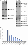
Immunoprecipitation of B17.2L and a subset of complex I subunits from normal human heart mitochondria. Mitochondria were isolated from normal human heart and immunoprecipitated with an immobilized anti-B17.2L antibody. (A) The immunoprecipitate was probed for the presence of B17.2L and the specific complex I subunits indicated to the right of the gels. COX-1, a subunit of complex IV, and SD70, a subunit of complex II, were used to control for nonspecific interactions. Lane I shows a sample of the mitochondrial protein applied to the immobilized antibody. Lane E represents half of the sample obtained following elution of the protein that specifically bound to the immobilized antibody. Lane W shows a sample of the final wash in the same volume as the eluate. (B) Quantitative analysis of the proteins immunoprecipitated by the anti-B17.2L antibody as obtained by densitometric analysis of the immunoblots.
Figure 8
Figure 8
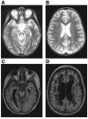
MRI of the brain of patient A. Transverse T2 weighted images taken at 3 years, 4 months of age (A and B). Transverse Fluid Attenuated Inversion Recovery (FLAIR) images taken at 10 years, 3 months of age (C and D).

Comment in

References

    1. Friedrich T, Weiss H. Modular evolution of the respiratory NADH:ubiquinone oxidoreductase and the origin of its modules. J. Theor. Biol. 1997;187:529–540. - PubMed
    1. Abdrakhmanova A, et al. Subunit composition of mitochondrial complex I from the yeast Yarrowia lipolytica. . Biochim. Biophys. Acta. 2004; 1658:148–156. - PubMed
    1. Carroll J, Fearnley IM, Shannon RJ, Hirst J, Walker JE. Analysis of the subunit composition of complex I from bovine heart mitochondria. Mol. Cell. Proteomics. 2003;2:117–126. - PubMed
    1. Gabaldon T, Rainey D, Huynen MA. Tracing the evolution of a large protein complex in the eukaryotes, NADH:ubiquinone oxidoreductase (complex I) J. Mol. Biol. 2005;348:857–870. - PubMed
    1. Grigorieff N. Three-dimensional structure of bovine NADH:ubiquinone oxidoreductase (complex I) at 22 A in ice. J. Mol. Biol. 1998;277:1033–1046. - PubMed

Publication types

MeSH terms