. 2005 Sep;1(1):e2.
doi: 10.1371/journal.ppat.0010002. Epub 2005 Sep 30.

Interaction between polyketide synthase and transporter suggests coupled synthesis and export of virulence lipid in M. tuberculosis

Affiliations

Interaction between polyketide synthase and transporter suggests coupled synthesis and export of virulence lipid in M. tuberculosis

Madhulika Jain et al. PLoS Pathog. 2005 Sep.

Abstract

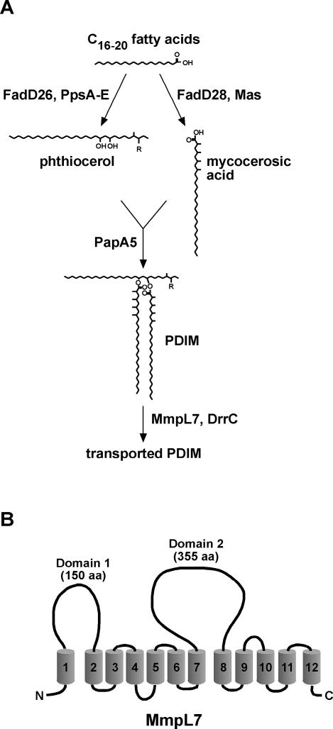
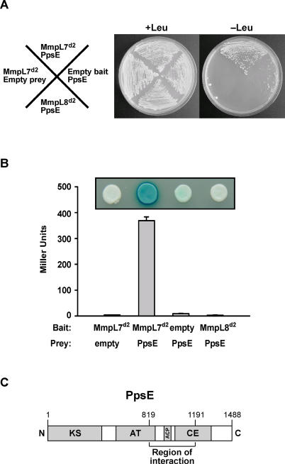
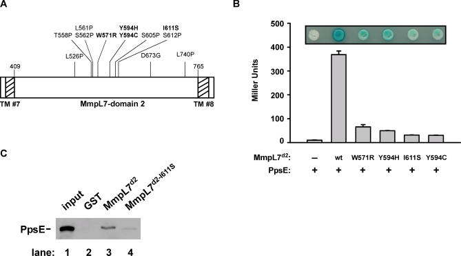
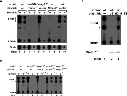
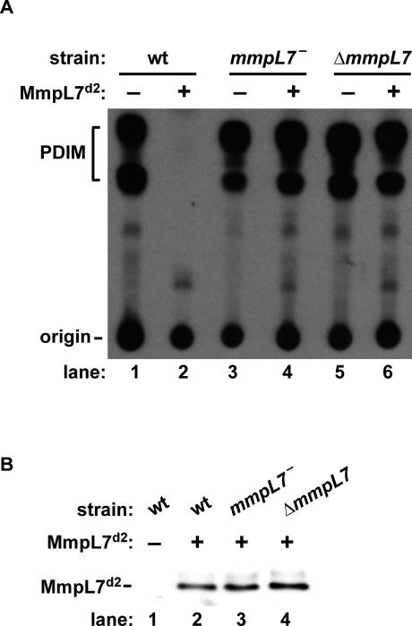
Virulent mycobacteria utilize surface-exposed polyketides to interact with host cells, but the mechanism by which these hydrophobic molecules are transported across the cell envelope to the surface of the bacteria is poorly understood. Phthiocerol dimycocerosate (PDIM), a surface-exposed polyketide lipid necessary for Mycobacterium tuberculosis virulence, is the product of several polyketide synthases including PpsE. Transport of PDIM requires MmpL7, a member of the MmpL family of RND permeases. Here we show that a domain of MmpL7 biochemically interacts with PpsE, the first report of an interaction between a biosynthetic enzyme and its cognate transporter. Overexpression of the interaction domain of MmpL7 acts as a dominant negative to PDIM synthesis by poisoning the interaction between synthase and transporter. This suggests that MmpL7 acts in complex with the synthesis machinery to efficiently transport PDIM across the cell membrane. Coordination of synthesis and transport may not only be a feature of MmpL-mediated transport in M. tuberculosis, but may also represent a general mechanism of polyketide export in many different microorganisms.


Conflict of interest statement

Competing interests. The authors have declared that no competing interests exist.

Figures

Figure 1
Figure 1. PDIM Synthesis and Export Pathway and Topology of MmpL7
(A) Schematic of the known steps in the PDIM synthesis and transport pathway. PpsA–E and Mas are polyketide synthases that extend fatty acids to phthiocerol and mycocerosic acids, respectively [16,17]. These are then esterified to produce PDIM. MmpL7 and DrrC are required for the transport of PDIM to the cell surface [8,10]. R is =O (keto) or –OCH3 (methoxy). (B) Predicted membrane topology of MmpL7 indicating the two non-TM domains 1 and 2.
Figure 2
Figure 2. MmpL7 Domain 2 Interacts with the PDIM Synthesis Enzyme PpsE
(A) Yeast two-hybrid reporter strains harboring the indicated bait and prey plasmids were streaked onto solid media with or without leucine. Growth on leucine-negative plates indicates a positive interaction. MmpL7d2, MmpL7 domain 2; MmpL8d2, MmpL8 domain 2. (B) The same strains as in (A) were transferred onto X-gal-containing indicator plates (inset), and reporter activity was quantified from liquid cultures using β-galactosidase assays. (C) Linear representation of full-length PpsE protein (1,488 amino acids) with the MmpL7 interaction region denoted. Catalytic domains of PpsE are also shown. ACP, acyl carrier protein; AT, acyl transferase; CE, condensing enzyme; KS, ketoacyl synthase.
Figure 3
Figure 3. Identification of Residues in MmpL7 Domain 2 Required for PpsE Interaction
(A) Twelve MmpL7 domain 2 mutants defective for PpsE binding were isolated in a reverse two-hybrid screen. These amino acid substitutions are displayed on a linear map of MmpL7 domain 2, with changes to amino acids other than proline or glycine in bold. Amino acid numbers correspond to positions in full-length MmpL7. TM domains 7 and 8 are denoted by hatched bars. (B) Yeast strains expressing the PpsE prey construct and various MmpL7 domain 2 bait plasmids were transferred onto X-gal indicator plates (inset), and reporter activity was quantified from liquid cultures by monitoring β-galactosidase activity. (C) Beads containing equal amounts of MmpL7 domain 2 and the I611S mutant were incubated with protein extracts containing myc-tagged PpsE and washed. Bound proteins were eluted and separated by SDS-PAGE, and PpsE was visualized by Western blot using anti-myc antibodies. GST-coated beads served as a negative control, and 1% of the protein extract added to the pulldown was loaded as a positive control (“input”).
Figure 4
Figure 4. MmpL7 Domain 2 Acts as a Dominant Negative Inhibitor of PDIM Synthesis In Vivo
(A) The indicated strains carrying either no plasmid (−), control vector (vector), or a plasmid with MmpL7 domain 2 under the control of the constitutive groEL2 promoter (MmpL7d2) were labeled with 14C-propionate. Surface-exposed lipids (S) were extracted by resuspension in hexanes, and cell pellets (P) were harvested by centrifugation. Lipids from both fractions were extracted and separated by TLC under solvent conditions to separate PDIM (upper panel, keto and methoxy forms) and SL-1 (lower panel). (B) Top: lipids were extracted as in (A) from pellets of wild-type cells carrying either no plasmid (−), the MmpL7 domain 2 expression plasmid (d2), or the MmpL7 domain 2 expression plasmid with the I611S mutation (d2-I611S). Bottom: Western blot analysis was performed to confirm equivalent expression of wild-type MmpL7 domain 2 and the I611S mutant by using antibodies against the hemagglutinin epitope tag. (C) Complementation of an mmpL7 M. tuberculosis strain with the wild-type (mmpL7wt) or the I611S mutant mmpL7 (mmpL7I611S). Surface-exposed lipids (S) and lipids associated with the remaining cell pellet (P) were extracted and separated by TLC to resolve PDIM as in (A).
Figure 5
Figure 5. Dominant Negative Effect of MmpL7 Domain 2 Requires the Presence of Full-Length MmpL7
(A) Wild-type cells, an mmpL7 transposon mutant (mmpL7 −), and a complete mmpL7 knockout (ΔmmpL7) carrying either no plasmid (−) or the MmpL7 domain 2 expression construct (+). Labeled lipids were extracted from pellets as described in Figure 4 and separated by TLC to resolve PDIM. (B) Western blot of MmpL7 domain 2 showing that it is expressed at equivalent levels in the different M. tuberculosis strains.
Figure 6
Figure 6. Model of PDIM Synthesis and Transport
MmpL7 interacts with PpsE, a subunit of the Pps enzyme required for PDIM synthesis. We propose that MmpL7 acts as a scaffold to recruit PDIM synthesis machinery, including Pps and perhaps Mas, leading to coordinate synthesis and transport of PDIM across the cytoplasmic membrane (CM). Whether MmpL7, or other factors, are required for delivery of PDIM through the peptidoglycan (PG) and mycolyl-arabinogalactan (mAG) layers is unclear.

References

    1. McKinney JD, Jacobs WR, Jr, Bloom BR. Persisting problems in tuberculosis. In: Fauci A, Krause R, editors. Emerging infections. London: Academic Press; 1998. pp. 51–146.
    1. Goren MB, Brokl O, Schaefer WB. Lipids of putative relevance to virulence in Mycobacterium tuberculosis: Phthiocerol dimycocerosate and the attenuation indicator lipid. Infect Immun. 1974;9:150–158. - PMC - PubMed
    1. Chan J, Fan XD, Hunter SW, Brennan PJ, Bloom BR. Lipoarabinomannan, a possible virulence factor involved in persistence of Mycobacterium tuberculosis within macrophages. Infect Immun. 1991;59:1755–1761. - PMC - PubMed
    1. Brennan PJ, Nikaido H. The envelope of mycobacteria. Annu Rev Biochem. 1995;64:29–63. - PubMed
    1. Ng V, Zanazzi G, Timpl R, Talts JF, Salzer JL, et al. Role of the cell wall phenolic glycolipid-1 in the peripheral nerve predilection of Mycobacterium leprae . Cell. 2000;103:511–524. - PubMed