. 2005 Sep;1(1):e4.
doi: 10.1371/journal.ppat.0010004. Epub 2005 Sep 30.

Influenza virus PB1-F2 protein induces cell death through mitochondrial ANT3 and VDAC1

Affiliations

Influenza virus PB1-F2 protein induces cell death through mitochondrial ANT3 and VDAC1

Dmitriy Zamarin et al. PLoS Pathog. 2005 Sep.

Abstract

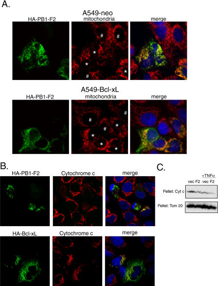
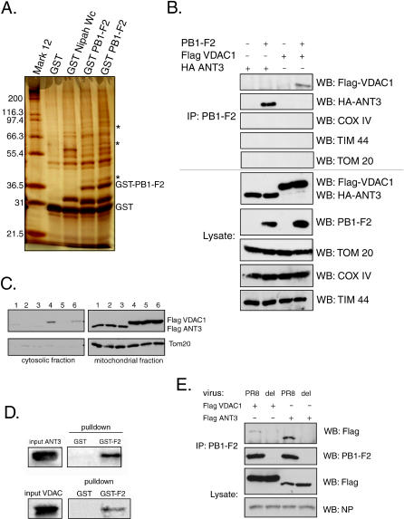
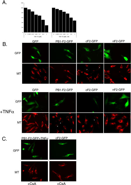
The influenza virus PB1-F2 is an 87-amino acid mitochondrial protein that previously has been shown to induce cell death, although the mechanism of apoptosis induction has remained unclear. In the process of characterizing its mechanism of action we found that the viral PB1-F2 protein sensitizes cells to apoptotic stimuli such as tumor necrosis factor alpha, as demonstrated by increased cleavage of caspase 3 substrates in PB1-F2-expressing cells. Moreover, treatment of purified mouse liver mitochondria with recombinant PB1-F2 protein resulted in cytochrome c release, loss of the mitochondrial membrane potential, and enhancement of tBid-induced mitochondrial permeabilization, suggesting a possible mechanism for the observed cellular sensitization to apoptosis. Using glutathione-S-transferase pulldowns with subsequent mass spectrometric analysis, we identified the mitochondrial interactors of the PB1-F2 protein and showed that the viral protein uniquely interacts with the inner mitochondrial membrane adenine nucleotide translocator 3 and the outer mitochondrial membrane voltage-dependent anion channel 1, both of which are implicated in the mitochondrial permeability transition during apoptosis. Consistent with this interaction, blockers of the permeability transition pore complex (PTPC) inhibited PB1-F2-induced mitochondrial permeabilization. Based on our findings, we propose a model whereby the proapoptotic PB1-F2 protein acts through the mitochondrial PTPC and may play a role in the down-regulation of the host immune response to infection.


Conflict of interest statement

Competing interests. The authors have declared that no competing interests exist.

Figures

Figure 1
Figure 1. PB1-F2 Protein Sensitizes Cells to Apoptosis in Response to Cytotoxic Stimuli
(A) Expression of PB1-F2 protein enhanced cleavage of poly-A ribose polymerase (PARP) in response to cytotoxic agents. 293T cells were transfected with either PB1-F2 (F2) or influenza virus NP (NP) for 24 h and were subsequently treated for 6 h with 50 ng/ml TNFα, 5 ng/ml TNF-related apoptosis-inducing ligand, 100 μM cisplatin, or UV-irradiation at 60 J/m2 as indicated. All cells were collected 6 h later, lysed, and processed by immunoblotting for cleaved PARP. (B) PB1-F2 protein predisposes cells to death by anoikis. A549 cells were transfected either with empty vector or with PB1-F2 and trypsinized 24 h post-transfection. Cells were incubated in PBS with 0.3% BSA for 15 min, pipetted onto microscope slides, and changes in cell morphology (membrane blebbing indicated by arrows) were visualized by phase-contrast microscopy [22]. (C) A subset of PB1-F2-expressing cells undergoes apoptosis. A549 cells were transfected with HA-tagged PB1-F2 for 24 h and stained for HA epitope and cleaved cytokeratin (M30).
Figure 2
Figure 2. PB1-F2 Protein-Mediated Enhancement of TNFα-Induced Apoptosis Is Inhibited by Bcl-xL
A549 cells containing a stably-integrated neomycin resistance gene (A549-neo) (A), or A549 cells stably overexpressing Bcl-xL (A549-Bcl-xL) (B) were transfected with either empty vector or vector encoding HA-tagged PB1-F2. At 24 h post-transfection, cells were treated with 50 ng/ml TNFα, where indicated. Then 6 h post-treatment, the cells were fixed and stained with M30 antibody to cleaved cytokeratin and with anti-HA antibody.
Figure 3
Figure 3. Influenza Virus PB1-F2 Protein Disrupts Mitochondria
(A) PB1-F2 disrupts reticulotubular mitochondrial organization. A549-neo and A549-Bcl-xL cells were transfected with HA-tagged PB1-F2 for 24 h and stained with antibody against HA tag and with human anti-mitochondrial serum as a marker for mitochondria. The cells with punctiform mitochondria are indicated by (*), while the cells with normal reticulotubular mitochondrial organization are indicated by (#). (B) PB1-F2 protein induces release of mitochondrial cytochrome c in transfected cells. HeLa cells were transfected with HA-tagged PB1-F2 for 24 h, treated with 50 ng/ml TNFα for 8 h, and stained with anti-HA antibody (green, secondary antibody FITC), anti-cytochrome c antibody (red, secondary antibody Alexa 568), and DAPI (blue). The cells were visualized by confocal microscopy. (C) Subcellular fractionation of 293T cells transfected with PB1-F2. 293T cells were transfected for 24 h and subsequently were either mock-treated or treated with 50 ng/ml TNFα. Cells were subsequently collected and fractionated into the cytosolic and mitochondrial fractions. The amount of cytochrome c remaining in the mitochondrial fraction was detected by Western blot. Tom20 protein was used as a loading control.
Figure 4
Figure 4. Influenza Virus PB1-F2 Protein Directly Permeabilizes Mitochondria
(A) Recombinant PB1-F2 protein caused release of cytochrome c from purified mouse mitochondria. Mouse mitochondria (50 μg total protein) were incubated for 1 h at 30 °C with recombinant protein of interest at indicated concentrations. The amount of cytochrome c released in the supernatant was assayed by immunoblotting using anti-cytochrome c antibody. Recombinant tBid protein was used as a control. The mitochondrial pellet was assayed for the Tom20 protein to ensure an equal amount of mitochondria was used. (B) PB1-F2 and tBid cause dissipation of the mitochondrial membrane potential. Mouse mitochondria (50 μg) were incubated for 10 min at 30 °C with the protein of interest at indicated concentrations. Relative loss in membrane potential was measured by uptake of membrane potential-sensitive JC-1 dye. Data is presented as percent fluorescence relative to mock-treated mitochondria. (C) PB1-F2 protein enhances tBid-induced cytochrome c release. Purified mouse mitochondria (50 μg) were incubated with 10 nM PB1-F2 or mock-treated for 15 min in 20 μl and were subsequently treated with recombinant tBid in the indicated concentrations for 1 h in a total volume of 30 μl. The supernatants were assayed for cytochrome c release by Western blot. The mitochondrial pellet was processed for Tom20 to ensure equal amount of mitochondria used.
Figure 5
Figure 5. PB1-F2 Protein Interacts with ANT3 and VDAC1
(A) Cellular proteins pulled down with GST-PB1-F2 were separated by 12% SDS-PAGE and silver-stained. The asterisks mark the protein bands that are unique to the GST-PB1-F2 lanes (~36 kDa, 55 kDa, and 80 kDa). Lanes 4 and 5 represent the results of two separate pulldown experiments. (B) PB1-F2 specifically interacts with ANT3 and VDAC1 but not other outer and inner mitochondrial membrane proteins. Interaction of PB1-F2 with ANT3 and VDAC1 was confirmed in 293T cells by coimmunoprecipitation with transfected HA-tagged ANT3 and flag-tagged VDAC1 (top five images). Tom20, COXIV, and TIM 44 proteins were used as the mitochondrial coimmunoprecipitation controls. (C) Transfected ANT3 and VDAC1 localize to mitochondria. 293T cells were transfected with Flag-tagged ANT3 and VDAC1 for 24 h and were subsequently fractionated to generate the cytosolic and mitochondrial fractions. Tom20 protein served as a control for the mitochondrial fraction. Each sample was processed in triplicate (lanes 1, 2, and 3, ANT3; lanes 4, 5, and 6, VDAC1). (D) PB1-F2 directly interacts with ANT3 and VDAC1. 35S-labeled ANT3 and VDAC1 were expressed in vitro using a rabbit reticulocyte lysate system and subjected to pulldown with either 5 μg GST (left lane) or GST-PB1-F2 (right lane). Proteins were separated by 12% SDS-PAGE and visualized by autoradiography. (E) PB1-F2 expressed during viral infection interacts with ANT3 and VDAC1. 293T cells were transfected with Flag-tagged ANT3 and VDAC1 proteins and 24 h later were infected with wild-type PR8 virus (PR8) or the virus knocked out for PB1-F2 expression (del) at an MOI of 2. 15 h after infection, the cells were collected and the immunoprecipitation with anti-PB1-F2 polyclonal serum was performed. Influenza virus NP was used as a control to show equal levels of infection between the samples. WB, Western blot.
Figure 6
Figure 6. Identification of PB1-F2 Protein Domains Responsible for Interaction with ANT3 and VDAC1
(A) PB1-F2 N- and C-terminal domains were cloned separately as N-terminal HA- and C-terminal GFP fusion proteins as follows. (1) GFP control; (2) full-length fusion protein (HA-PB1-F2-GFP); (3) C-terminal 38–87-amino acid domain fusion protein (HA-cF2-GFP); (4) N-terminal 1–37 amino acid domain fusion protein (HA-nF2-GFP); (5) N-terminal domain fusion protein with cytochrome oxidase mitochondrial targeting sequence (HA-MTS-nF2-GFP); and (6) control MTS-GFP fusion protein (HA-MTS-GFP). (B) Localization of fusion proteins was determined in transfected HeLa cells (green, GFP-fusion protein; red, Mitotracker dye staining for mitochondria; blue, DAPI nuclear stain). (C and D) Interaction of fusion proteins with ANT3 and VDAC1 was determined by cotransfecting 293T cells with each fusion construct and a vector encoding Flag-tagged ANT3 or VDAC1, respectively. Immunoprecipitation was performed with an anti-HA antibody, with subsequent immunoblotting for Flag-tagged ANT3 or VDAC1.
Figure 7
Figure 7. PB1-F2 Protein Induces Mitochondrial Permeabilization in ANT3-Dependent Manner
(A) Recombinant PB1-F2 protein was incubated with 50 μg of purified mouse liver mitochondria in the presence or absence of 50 μM BA for 30 min. The mitochondria were further processed for assessment of membrane potential by JC-1 fluorescence at 590 nm. (B) PB1-F2 induces loss of mitochondrial membrane potential in transfected cells. HeLa cells were transfected with GFP fusion constructs of PB1-F2, and 12 h later were treated with 50 ng/ml TNFα for 8 h, where indicated. Cells were subsequently stained with Mitotracker CMXRos Red dye. Cells with dissipated membrane potential are indicated by (*). (C) The mitochondrial permeability transition inhibitor CsA inhibits PB1-F2-induced loss of mitochondrial membrane potential. HeLa cells in presence of CsA were transfected and treated as in (B) and stained with Mitotracker dye.
Figure 8
Figure 8. PB1-F2 Induces Apoptosis Acting through Components of the PTPC
(A) ANT3 blocker BA inhibits PB1-F2-mediated sensitization of cells to apoptosis. A549 cells were transfected with PB1-F2 for 20 h and then treated with TNFα as indicated, either in the presence or absence of 50 μM BA. The cells were collected 8 h later and stained with M30 antibody to cleaved cytokeratin. (B) Proposed models of PB1-F2 action during infection. During early stages of the infection, PB1-F2 localizes to mitochondria, where it interacts with ANT3 and VDAC and predisposes the mitochondria to permeability transition. Later in the infection, when more PB1-F2 is synthesized, and upon induction of antiviral apoptotic signaling pathways, the mitochondria undergo the permeability transition, which results in the induction of apoptosis. PB1-F2-induced mitochondrial permeabilization can proceed through three possible mechanisms, as indicated on the right graphic: (1) enhancement of the pore complex formation through direct interaction with ANT3 and VDAC1; (2) independent permeabilization of the inner and outer mitochondrial membranes with the help of ANT3 and VDAC1, respectively; and (3) direct permeabilization of the mitochondrial membranes.

References

    1. Garcia-Sastre A. Inhibition of interferon-mediated antiviral responses by influenza A viruses and other negative-strand RNA viruses. Virology. 2001;279:375–384. - PubMed
    1. Garcia-Sastre A, Egorov A, Matassov D, Brandt S, Levy DE, et al. Influenza A virus lacking the NS1 gene replicates in interferon-deficient systems. Virology. 1998;252:324–330. - PubMed
    1. Chen W, Calvo PA, Malide D, Gibbs J, Schubert U, et al. A novel influenza A virus mitochondrial protein that induces cell death. Nat Med. 2001;7:1306–1312. - PubMed
    1. Gibbs JS, Malide D, Hornung F, Bennink JR, Yewdell JW. The influenza A virus PB1-F2 protein targets the inner mitochondrial membrane via a predicted basic amphipathic helix that disrupts mitochondrial function. J Virol. 2003;77:7214–7224. - PMC - PubMed
    1. Yamada H, Chounan R, Higashi Y, Kurihara N, Kido H. Mitochondrial targeting sequence of the influenza A virus PB1-F2 protein and its function in mitochondria. FEBS Lett. 2004;578:331–336. - PubMed