. 2005 Sep;1(1):e7.
doi: 10.1371/journal.ppat.0010007. Epub 2005 Sep 30.

Inhibition of MHC class I is a virulence factor in herpes simplex virus infection of mice

Affiliations

Inhibition of MHC class I is a virulence factor in herpes simplex virus infection of mice

Mark T Orr et al. PLoS Pathog. 2005 Sep.

Abstract

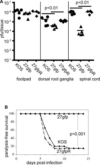
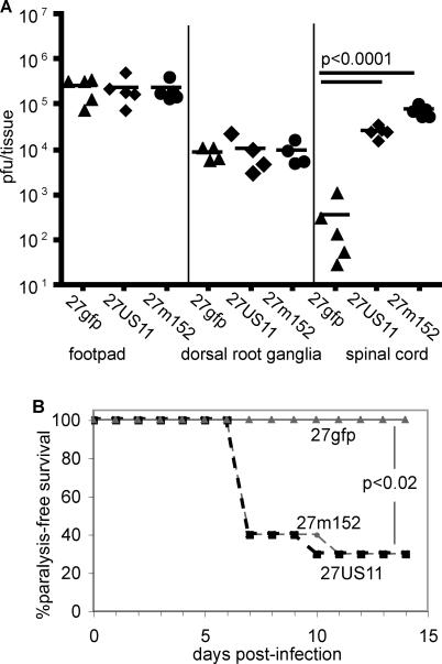
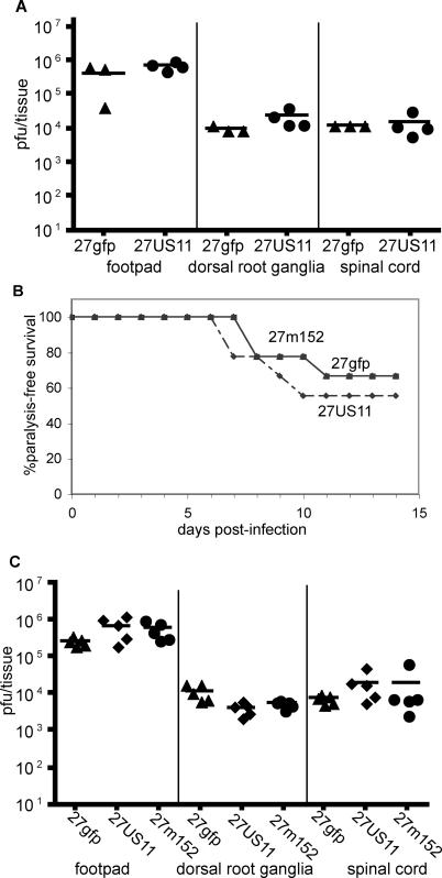
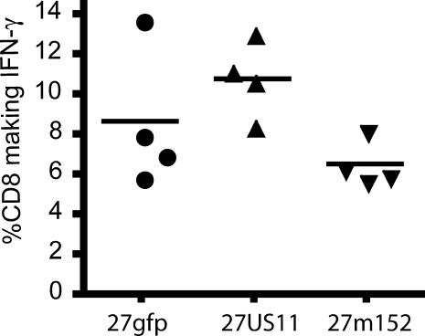
Herpes simplex virus (HSV) has a number of genes devoted to immune evasion. One such gene, ICP47, binds to the transporter associated with antigen presentation (TAP) 1/2 thereby preventing transport of viral peptides into the endoplasmic reticulum, loading of peptides onto nascent major histocompatibility complex (MHC) class I molecules, and presentation of peptides to CD8 T cells. However, ICP47 binds poorly to murine TAP1/2 and so inhibits antigen presentation by MHC class I in mice much less efficiently than in humans, limiting the utility of murine models to address the importance of MHC class I inhibition in HSV immunopathogenesis. To address this limitation, we generated recombinant HSVs that efficiently inhibit antigen presentation by murine MHC class I. These recombinant viruses prevented cytotoxic T lymphocyte killing of infected cells in vitro, replicated to higher titers in the central nervous system, and induced paralysis more frequently than control HSV. This increase in virulence was due to inhibition of antigen presentation to CD8 T cells, since these differences were not evident in MHC class I-deficient mice or in mice in which CD8 T cells were depleted. Inhibition of MHC class I by the recombinant viruses did not impair the induction of the HSV-specific CD8 T-cell response, indicating that cross-presentation is the principal mechanism by which HSV-specific CD8 T cells are induced. This inhibition in turn facilitates greater viral entry, replication, and/or survival in the central nervous system, leading to an increased incidence of paralysis.


Conflict of interest statement

Competing interests. The authors have declared that no competing interests exist.

Figures

Figure 1
Figure 1. Generation of rHSVs
(A) rHSVs expressing MCMV m152 (27m152), HCMV US11 (27US11), or only the selection cassette (27gfp) were generated via homologous recombination with KOS strain HSV-1. Contents and location of insertions are indicated. Arrows indicate direction of transcription. The probe used to isolate correctly recombined viruses is indicated. (B) Genomic DNA from KOS, 27US11, 27m152, 27gfp, and 27gfpR was digested with the indicated restriction enzymes and probed for the junction of HSV UL26–UL27. The predicted band sizes are indicated in the tabular inset.
Figure 2
Figure 2. 27US11 and 27m152 Inhibit Murine MHC Class I Preventing Lysis by HSV-Specific CTL
(A) K-BALB (H-2d) and (B) MC57G (H-2b) fibroblast cell lines were uninfected (solid line) or infected (dashed line) at an MOI of 5:1 with 27gfp, 27US11, or 27m152 for 18 h and analyzed for surface MHC class I expression. Filled histograms are isotype controls. The percentage reduction in mean fluorescent intensity from uninfected to infected cells is indicated. (C) and (D) Cells were infected as for (A) and (B) and co-incubated with CTL isolated from HSV-infected (C) BALB/c or (D) BALB.B mice at the indicated effector-to-target ratio.
Figure 3
Figure 3. 27m152, but Not 27US11, Evades NK Recognition by Inhibiting NKG2D Ligands
(A) BALB/c fibroblasts were uninfected (solid line) or infected (dashed line) for 18 h at an MOI of 5:1 with 27gfp, 27US11, or 27m152 (dashed line) and stained with NKG2D tetramer. Solid histogram is uninfected cells stained with an irrelevant tetramer. The percentage reduction in mean fluorescent intensity from uninfected to infected cells is indicated. (B) Cells were prepared as in (A) and co-incubated with splenocytes from naïve RAG1−/− BALB/c mice treated with polyI:C 24 h earlier at the indicated effector-to-target ratio.
Figure 4
Figure 4. Single-Step Growth Kinetics of rHSVs Are Similar to KOS
Vero cells were infected at an MOI of 5:1 with KOS plus (A) 27m152 or 27US11, or (B) 27gfp. Cells and supernatants were harvested at indicated times and viral titers were determined on vero cells.
Figure 5
Figure 5. The Selection Cassette Attenuates Neuroinvasiveness and Neurovirulence
BALB/c mice were infected in the hind footpads with 2.5 × 10⁵ pfu of KOS, 27gfp, or 27gfpR. (A) The indicated tissues were isolated on day 6, homogenized, and viral titers were determined on vero cells. (B) Ten (27gfp) or 20 (KOS and 27gfpR) mice per virus were monitored for paralysis induction for 14 d. Mice displaying ataxia or paralysis were euthanized.
Figure 6
Figure 6. Inhibition of MHC Class I Increases Neuroinvasiveness and Neurovirulence
BALB/c were infected in the hind footpads with 2.5 × 10⁵ pfu of 27gfp, 27US11, or 27m152. (A) The indicated tissues were isolated on day 6, homogenized, and viral titers were determined on vero cells. (B) Ten mice per virus were monitored for paralysis induction for 14 d. Mice displaying ataxia or paralysis were euthanized.
Figure 7
Figure 7. Differences in Neuroinvasiveness and Neurovirulence Are Dependent on MHC Class I and CD8 T Cells
β2m−/− BALB/c mice were infected in the hind footpads with 3.0 × 10⁴ pfu of 27gfp, 27US11, or 27m152. Note that a lower inoculum was used in these experiments with β2m−/− mice than in wild-type BALB/c mice shown in other figures. (A) The indicated tissues were isolated on day 6, homogenized, and viral titers were determined on vero cells. (B) Nine mice per virus were monitored for paralysis induction for 14 d. Mice displaying ataxia or paralysis were euthanized. (C) BALB/c mice were depleted of CD8 cells and infected with 2.5 × 10⁵ pfu of the indicated virus in the hind footpads. Viral titers were determined on day 6.
Figure 8
Figure 8. The Size of the CD8 T-Cell Response to rHSVs Is Not Altered by MHC Class I Inhibition
Lymphocytes from the draining popliteal lymph nodes were isolated from BALB.B mice on day 6 of infection with 27gfp, 27US11, or 27m152. Lymphocytes were stimulated with HSV gB498–505 for 5 h then stained for CD8 and intracellular IFN-γ. Of the unstimulated CD8+ cells, <0.1% were IFN-γ+.

Similar articles

Cited by

References

    1. Alcami A, Koszinowski UH. Viral mechanisms of immune evasion. Immunol Today. 2000;21:447–455. - PMC - PubMed
    1. Tortorella D, Gewurz BE, Furman MH, Schust DJ, Ploegh HL. Viral subversion of the immune system. Annu Rev Immunol. 2000;18:861–926. - PubMed
    1. Fruh K, Gruhler A, Krishna RM, Schoenhals GJ. A comparison of viral immune escape strategies targeting the MHC class I assembly pathway. Immunol Rev. 1999;168:157–166. - PubMed
    1. Fruh K, Ahn K, Peterson PA. Inhibition of MHC class I antigen presentation by viral proteins. J Mol Med. 1997;75:18–27. - PubMed
    1. Yewdell JW, Hill AB. Viral interference with antigen presentation. Nat Immunol. 2002;3:1019–1025. - PubMed