. 2006 Jan 1;393(Pt 1):381-7.
doi: 10.1042/BJ20051133.

Physical and functional interactions between Escherichia coli MutY and endonuclease VIII

Affiliations

Physical and functional interactions between Escherichia coli MutY and endonuclease VIII

A-Lien Lu et al. Biochem J. .

Abstract

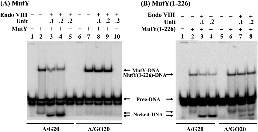
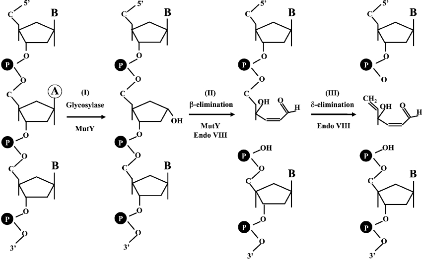
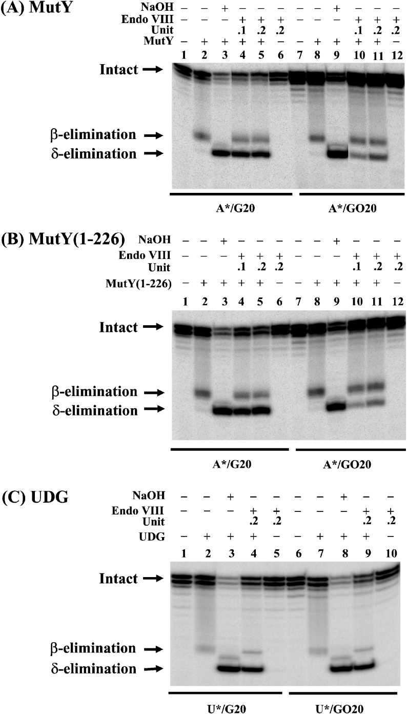
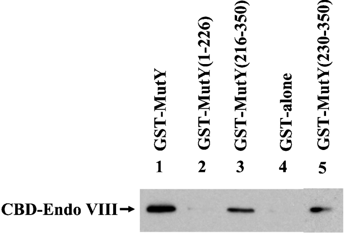
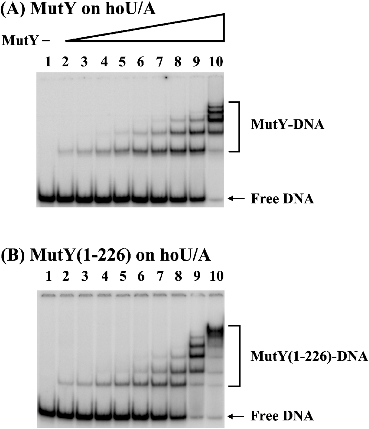
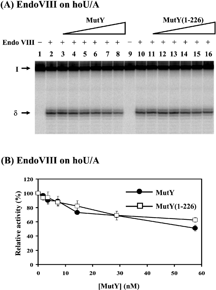
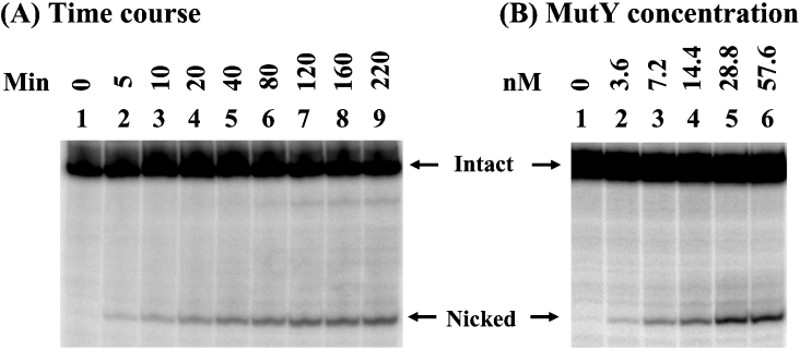
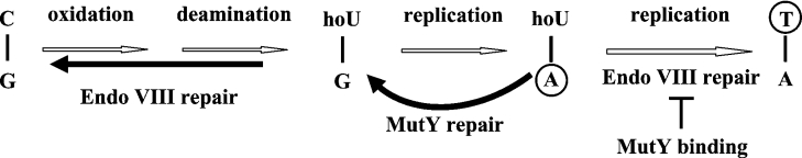
Both GO (7,8-dihydro-8-oxoguanine) and hoU (5-hydroxyuracil) are highly mutagenic because DNA polymerase frequently misincorporates adenine opposite these damaged bases. In Escherichia coli, MutY DNA glycosylase can remove misincorporated adenine opposite G or GO on the template strand during DNA replication. MutY remains bound to the product that contains an AP (apurinic/apyrimidinic) site. Endo VIII (endonuclease VIII) can remove oxidized pyrimidine and weakly remove GO by its DNA glycosylase and beta/delta-elimination activities. In the present paper, we demonstrate that Endo VIII can promote MutY dissociation from AP/G, but not from AP/GO, and can promote beta/delta-elimination on the products of MutY. MutY interacts physically with Endo VIII through its C-terminal domain. MutY has a moderate affinity for DNA containing a hoU/A mismatch, which is a substrate of Endo VIII. MutY competes with Endo VIII and inhibits Endo VIII activity on DNA that contains a hoU/A mismatch. Moreover, MutY has a weak adenine glycosylase activity on hoU/A mismatches. These results suggest that MutY may have some role in reducing the mutagenic effects of hoU.


Figures

Figure 1
Figure 1. Effects of Endo VIII on DNA binding by MutY and MutY-(1–226)
(A) Effects of Endo VIII on DNA-binding activity of MutY. Lane 1, 5′-end-labelled A/G-containing 20-mer DNA (A/G20). Lane 2, 1.8 fmol (90 pM) of A/G20 DNA substrate was incubated with MutY (1.8 nM). Lanes 3 and 4, same as lane 2, but with 0.1 unit (0.17 nM) and 0.2 unit (0.34 nM) of Endo VIII respectively. Lane 5, 1.8 fmol (90 pM) of A/G20 DNA substrate was incubated with Endo VIII (0.2 unit or 0.34 nM). Lanes 6–10 are similar to lanes 1–5, except using A/GO-containing 20-mer DNA (A/GO20) and the MutY concentration is 0.11 nM. All reaction mixtures were incubated for 30 min at 37 °C and fractionated on a 6% native gel. (B) Effects of Endo VIII on DNA-binding activity of MutY-(1–226). Lanes 1–4 are similar to lanes 1–4 in (A) and lanes 5–8 are similar to lanes 6–9 in (A), except that MutY-(1–226) was used. Free DNA, nicked DNA, MutY–DNA complex and MutY-(1–226)–DNA complex are indicated.
Figure 2
Figure 2. Reactions of MutY DNA glycosylase and β/δ-elimination activities of Endo VIII
Step I, MutY DNA glycosylase removes a mismatched adenine (circled) which is paired with GO, guanine or cytosine on the other strand (not shown). The MutY product contains an AP site. Step II, the weak AP lyase activity of MutY and the β-elimination activity of Endo VIII cleave the DNA backbone to generate two fragments, one of which contains an unsaturated sugar moiety. Step III, the δ-elimination activity of Endo VIII removes the unsaturated sugar to produce a DNA fragment with a 3′ phosphate group.
Figure 3
Figure 3. Endo VIII activity on the DNA products generated by MutY, MutY-(1–226) and UDG
(A) The activity of Endo VIII on DNA products of MutY. Lanes 1 and 7, 5′-end-labelled A/G- and A/GO-containing DNA respectively. A/G20 DNA substrate (1.8 fmol or 180 pM) was incubated with 7.2 nM MutY (lanes 2–5) and A/GO20 DNA substrate (1.8 fmol or 180 pM) was incubated with 1.8 nM MutY (lanes 8–11) at 37 °C for 30 min. Endo VIII [0.1 unit (0.34 nM) or 0.2 unit (0.68 nM)] was added to reaction mixtures in lanes 4–6 and lanes 10–12 as indicated. Lanes 3 and 9, after glycosylase reaction, the mixtures were supplemented with 1 μl of 1 M NaOH and heated at 90 °C for 30 min. Formamide dye (5 μl) was added to the sample, which was heated at 90 °C for 2 min, and 5 μl of the mixture was loaded on to a 14% (w/v) polyacrylamide sequencing gel containing 7 M urea. The positions of the intact substrate and the products generated by β- and δ-eliminations are marked. (B) The activity of Endo VIII on DNA products of MutY-(1–226). Reactions were similar to (A) except that MutY-(1–226) was used. (C) The activity of Endo VIII on DNA products of UDG DNA glycosylase. Reactions were similar to (A), except 0.01 unit of UDG with U/G20 and U/GO20 DNA substrates were used. Asterisks mark the 32P-labelled DNA strands.
Figure 4
Figure 4. Physical interaction of MutY with Endo VIII
GST alone and several GST–MutY constructs (300 ng) immobilized on glutathione–Sepharose were incubated with 0.13 mg of E. coli extracts expressing CBD-tagged Endo VIII protein as described in the Materials and methods section. The pellets were fractionated on an SDS/10% polyacrylamide gel followed by Western blot analysis using the antibody against CBD. Lane 1, GST-tagged intact MutY (residues 1–350); lane 2, GST–MutY-(1–226); lane 3, GST–MutY-(216–350); lanes 4, GST alone; lane 5, GST–MutY-(230–350).
Figure 5
Figure 5. Binding activities of MutY and MutY-(1–226) with DNA containing hoU/A mismatch
(A) HoU/A-containing DNA was incubated with MutY at various protein concentrations at 37 °C for 30 min. Lane 1, DNA labelled at the 5′ end of the hoU-containing strand. Lanes 2–10, hoU/A-containing DNA was incubated with MutY protein at concentrations of 0.9, 1.8, 3.6, 7.2, 14.4, 28.8, 57.6, 115.2 or 230.4 nM respectively. Products were fractionated on 6% native gels. (B) is similar to (A), except using MutY-(1–226). The positions of the free DNA, MutY–DNA complex and MutY-(1–226)–DNA complex are marked.
Figure 6
Figure 6. MutY and MutY-(1–226) can inhibit Endo VIII activity
(A) Activity of Endo VIII on hoU/A substrate can be inhibited by MutY and MutY-(1–226). Lanes 1 and 9, hoU/A-containing DNA labelled at 5′ end of the hoU-containing strand. DNA substrate (1.8 fmol or 180 pM) was incubated with 0.34 nM (0.1 unit) of Endo VIII (lanes 2–8 and 10–16) at 37 °C for 30 min. MutY (1.8, 3.6, 7.2, 14.4, 28.8 or 57.6 nM) was added to reaction mixtures in lanes 3–8. MutY-(1–226) (1.8, 3.6, 7.2, 14.4, 28.8 or 57.6 nM) was added to reaction mixtures in lanes 11–16. After reactions, 5 μl of formamide dye was added to the sample, which was heated at 90 °C for 2 min, and 5 μl of the mixture was loaded on to a 14% (w/v) polyacrylamide sequencing gel containing 7 M urea. The positions of the intact substrate (I) and the products generated by δ-elimination (δ) are marked. (B) Quantitative analyses of MutY and MutY-(1–226) inhibition on Endo VIII activity on hoU/A substrate. Results are means±S.D. from PhosphorImager quantitative analyses of gel images from three experiments as represented in (A).
Figure 7
Figure 7. MutY glycosylase activity on DNA containing a hoU/A mismatch
(A) Time-course study of MutY glycosylase activity on hoU/A-containing DNA. DNA (1.8 fmol) labelled at the 5′ end of the adenine-containing strand was incubated with 14.4 nM MutY at 37 °C. (B) MutY glycosylase activity on hoU/A-containing DNA at different protein concentrations. hoU/A-containing DNA (1.8 fmol) labelled at the 5′ end of the adenine-containing strand was incubated with MutY protein at 3.6, 7.2, 14.4, 28.8 or 57.6 nM at 37 °C for 1 h. After incubation, the reaction mixtures were supplemented with 1 μl of 1 M NaOH and heated at 90 °C for 30 min. Formamide dye (5 μl) was added to the sample, which was heated at 90 °C for 2 min, and 5 μl of the mixture was loaded on to a 14% (w/v) polyacrylamide sequencing gel containing 7 M urea.
Figure 8
Figure 8. E. coli MutY may have a role in preventing the mutagenic effects of hoU
hoU in DNA can be derived from oxidation, deamination and dehydration of cytosine. The Endo VIII glycosylase/AP lyase removes hoU adducts. When hoU/G is not repaired by Endo VIII, adenines are frequently incorporated opposite hoU bases during DNA replication. A/hoU mismatches may be repaired to G/hoU by the MutY pathway. Without repair, the second round of DNA replication of A/hoU can generate G:C→A:T transitions. If Endo VIII repairs an A/hoU mismatch when hoU is on the parental DNA strand, a G:C→A:T mutation occurs. MutY may inhibit Endo VIII repair on A/hoU. Bases on the daughter DNA strands are circled. Repair pathways that can reduce mutagenesis are marked with solid thick lines.

Similar articles

Cited by

References

    1. Halliwell B., Gutteridge J. M. New York: Oxford University Press; 1989. Free Radicals in Biology and Medicine.
    1. Michaels M. L., Miller J. H. The GO system protects organisms from the mutagenic effect of the spontaneous lesion 8-hydroxyguanine (7,8-dihydro-8-oxoguanine) J. Bacteriol. 1992;174:6321–6325. - PMC - PubMed
    1. Tchou J., Grollman A. P. Repair of DNA containing the oxidatively-damaged base 8-hydroxyguanine. Mutat. Res. 1993;299:277–287. - PubMed
    1. Lu A.-L., Li X., Gu Y., Wright P. M., Chang D.-Y. Repair of oxidative DNA damage. Cell Biochem. Biophys. 2001;35:141–170. - PubMed
    1. Maki H., Sekiguchi M. MutT protein specifically hydrolyses a potent mutagenic substrate for DNA synthesis. Nature (London) 1992;355:273–275. - PubMed

Publication types

MeSH terms

LinkOut - more resources