. 2006 Jan 1;393(Pt 1):397-409.
doi: 10.1042/BJ20051030.

Nkx3.1 binds and negatively regulates the transcriptional activity of Sp-family members in prostate-derived cells

Affiliations

Nkx3.1 binds and negatively regulates the transcriptional activity of Sp-family members in prostate-derived cells

Steven O Simmons et al. Biochem J. .

Abstract

Nkx3.1 is a homeodomain-containing transcription factor that is expressed early in the development of the prostate gland and is believed to play an important role in the differentiation of prostatic epithelia. Loss of Nkx3.1 protein expression is often an early event in prostate tumorigenesis, and the abundance of Nkx3.1-negative epithelial cells increases with disease progression. In a number of systems, homeodomain proteins collaborate with zinc-finger-containing transcription factors to bind and regulate target genes. In the present paper, we report that Nkx3.1 collaborates with Sp-family members in the regulation of PSA (prostate-specific antigen) in prostate-derived cells. Nkx3.1 forms protein complexes with Sp proteins that are dependent on their respective DNA-binding domains and an N-terminal segment of Nkx3.1, and Nkx3.1 negatively regulates Sp-mediated transcription via Trichostatin A-sensitive and -insensitive mechanisms. A distal 1000 bp portion of the PSA promoter is required for transrepression by Nkx3.1, although Nkx3.1 DNA-binding activity is itself not required. We conclude that Nkx3.1 negatively regulates Sp-mediated transcription via the tethering of histone deacetylases and/or by inhibiting the association of Sp proteins with co-activators.


Figures

Figure 1
Figure 1. Transactivation of the human PSA promoter by Sp-family members and transrepression by mNkx3.1
(A) Effects of Sp-family members and Nkx3.1 on PSA transcription. Human DU145 prostate cells were transiently transfected with mNkx3.1 alone (500 ng; Nkx), with Sp1-5 alone (500 ng), or with mNkx3.1 (500 ng) and an Sp-family member (500 ng; denoted by Sp-family member+N, e.g. 1+N). Basal levels of PSA transcription were set equal to 1, and the mean fold changes in PSA transactivation normalized to an internal firefly luciferase control following 48 h of cultivation post-transfection are plotted. Empty expression vector DNA was included in control reactions to maintain constant input DNA concentrations (2 μg per plate). Results are means±S.D. for at least five independent plates of transfected cells. (B) Effects of Sp-family members and Nkx3.1 on PSA transcription following treatment with TSA. DU145 cells were transfected and processed as in (A), except that cells were treated with 100 nM TSA 24 h after transfection. Basal levels of PSA transcription were 10-fold greater than that shown in (A) and therefore PSA values were set equal to 10. Results are means±S.D. for at least five independent plates of transfected cells.
Figure 2
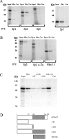
Figure 2. In vitro protein–protein-binding assay
(A) Specific binding of Nkx3.1 by Sp-family members. In vitro translated (IVT) radiolabelled Sp proteins were synthesized in reticulocyte lysates and incubated with a GST-fusion protein prepared using a full-length mNkx3.1 cDNA (Nkx) or a control fusion protein (GST–FSH; indicated by Con). In vitro translated proteins (10% of input) were resolved alone (indicated by Input) as controls. Molecular-mass markers (sizes in kDa) are indicated on the left. (B) The DNA-binding domain of Sp-family members is necessary and sufficient for complex formation with Nkx3.1. A full-length GST–mNkx3.1 fusion protein (Nkx), GST–FSH (indicated by Con) or a GST-fusion prepared from the Sp2 DNA-binding domain (pGEX1N-Sp2Zn; indicated by Zn) was challenged with in vitro translated radiolabelled Sp2 protein, an Sp2 derivative lacking the DNA-binding domain or mNkx3.1 respectively. In vitro translated (IVT) proteins (10% of input) were resolved alone as controls (indicated by Input), and radiolabelled proteins examined are indicated beneath the gel. Molecular-mass markers (sizes in kDa) are indicated on the left. (C) Two non-contiguous regions of Nkx3.1 carry binding sites for Sp-family members. In vitro translated radiolabelled Sp2 was synthesized in reticulocyte lysates and incubated with a full-length mNkx3.1 GST-fusion protein (Nkx) or derivatives carrying various portions of mNkx3.1 as depicted in (D) (indicated at the top of each lane). Sp2 (10% of input) was resolved as a control (indicated by Input), and GST–FSH was employed as a negative control (indicated by Con). Molecular-mass markers (sizes in kDa) are indicated on the left. (D) Schematic diagram of mNkx3.1 and truncated derivatives employed as GST-fusion proteins in (C). The homeodomain is indicated by a hatched box as are amino acid endpoints for Nkx3.1 deletions.
Figure 3
Figure 3. The DNA-binding domains of Nkx3.1 and Sp proteins form complexes in mammalian cells
(A) Schematic diagram of mammalian two-hybrid fusion proteins. The N-terminal 229 amino acids of Renilla luciferase were fused to the DNA-binding domain (amino acids 519–606) of Sp2, creating pNhRL-Zn. The C-terminal 82 amino acids of Renilla luciferase were fused to the human Nkx3.1 homeodomain (amino acids 124–183), creating pChRL-HD. An analogous construct carrying the human Nkx3.1 homeodomain upstream of the Renilla C-terminus created pHD-ChRL. The Sp2 DNA-binding domain is indicated by a hatched box, and the Nkx3.1 homeodomain is indicated by a stippled box. Amino acid endpoints are indicated above each schematic diagram. (B) Reconstitution of luciferase activity in vivo. COS-1 cells were transfected with 0.5 μg of pNhRL-ZN and 0.5 μg of pChRL, an empty control vector, or 0.5 μg of fusion constructs carrying the human Nkx3.1 homeodomain (pChRL-HD and pHD-ChRL). As an additional control, 0.5 μg of pChRL-HD was transfected with 0.5 μg of pNhRL, the corresponding empty control vector. An irrelevant plasmid was included in all transfections to maintain constant input DNA concentrations (2 μg per plate). Results are means±S.D. of Renilla luciferase activity for six or more plates of transfected cells. (C) Competition for reconstitution of Renilla luciferase activity by wild-type Sp-family members. COS-1 cells were transfected with 0.5 μg of pNhRL-ZN and 0.5 μg of pChRL-HD as well as increasing amounts (100–1000 ng) of expression vectors carrying wild-type Sp-family members or 1 μg of empty expression vector (pCMV4). Where appropriate, an irrelevant plasmid was included in transfections to maintain constant input DNA concentrations (2 μg per plate). Results are means±S.D. of Renilla luciferase activity for six or more plates of transfected cells.
Figure 4
Figure 4. Protein–DNA-binding assays with recombinant human Nkx3.1 protein
(A) Recombinant hNkx3.1 binds DNA specifically in vitro. Radiolabelled oligonucleotide probes carrying consensus (WT) or mutated (Mut) Nkx3.1-binding sites were incubated with or without various amounts of bacterially expressed hNkx3.1 as indicated. Protein–DNA complexes were challenged with anti-human (T-19 and N-15) or anti-mouse (L-15) Nkx3.1 antibodies where indicated. Nkx3.1 protein–DNA complexes are indicated by Nkx, antibody-bound super-shifted complexes are indicated by SS, and free probe is indicated by FP. (B) Competition experiment using wild-type and mutated oligonucleotides. A radiolabelled oligonucleotide probe carrying a consensus Nkx3.1-binding site was incubated alone or with 100 ng of recombinant hNkx3.1 protein and challenged with increasing amounts of unlabelled wild-type (WT) or mutated (Mut) oligonucleotides. Molar excesses of unlabelled competitor DNAs are indicated. Nkx3.1 protein–DNA complexes are indicated by Nkx, and free probe is indicated by FP.
Figure 5
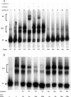
Figure 5. Protein–DNA-binding assays with recombinant human Nkx3.1 and Sp proteins
(A) Protein–DNA-binding assays with recombinant hNkx3.1 and human Sp-family members. A radiolabelled oligonucleotide probe (NkxSp-10) carrying consensus Nkx3.1- and Sp2-binding sites separated by 5 bp was incubated alone or with 100 ng of recombinant hNkx3.1 and baculovirus extracts carrying recombinant Sp1, Sp2 or Sp3 protein (proteins added are indicated by +). Nkx3.1 protein–DNA complexes are indicated by Nkx. Sp1, Sp2 and Sp3 indicate their respective protein–DNA complexes, and free probe is also indicated (FP). (B) Extended electrophoresis of protein–DNA-binding assays with recombinant hNkx3.1 and human Sp3 proteins. A radiolabelled NkxSp-5 probe was incubated with baculovirus extracts carrying recombinant Sp3 protein and 100 ng of recombinant hNkx3.1 (proteins added are indicated by +). Protein–DNA complexes were challenged with anti-hNkx3.1 antibody T-19 (where indicated by +). (C) Protein–DNA-binding assays with recombinant hNkx3.1 and human Sp-family members using a probe carrying a consensus Sp-binding site. A radiolabelled probe carrying a consensus Sp2-binding site was incubated alone or with baculovirus extracts carrying recombinant Sp1, Sp2 or Sp3 protein, and 100 ng of recombinant hNkx3.1 (proteins added are indicated by +). Protein–DNA complexes were challenged with anti-hNkx3.1 antibody T-19 (where indicated by +). Sp1, Sp2 and Sp3 indicate their respective protein–DNA complexes, and FP indicates free probe.
Figure 6
Figure 6. Sensitivity of PSA promoter deletion mutants to transactivation by Sp3 and transrepression by hNkx3.1
(A) Schematic diagram of human PSA promoter (5300 bp; drawn 5′→3′). Restriction sites used to generate deletion mutants are indicated as are predicted Nkx3.1- (stippled boxes) and Sp- (solid boxes) binding sites. Predicted protein-binding sites on the sense strand are shown above the line, and those on the antisense strand are shown below the line. (B) Transcriptional response of wild-type and deleted PSA promoter constructs following the ectopic expression of Sp3, hNkx3.1, or Sp3 and hNkx3.1 in DU145 cells. Results are changes±S.D. of mean fold transactivation of each construction relative to its basal activity (set equal to 1) for five independent plates of cells transfected with each construction.
Figure 7
Figure 7. Transcriptional regulation of wild-type and mutated PSA promoter constructs by Sp3 and wild-type Nkx3.1 or DNA binding-deficient homeodomain mutants
(A) DU145 cells were transfected with promoter constructs alone, in conjunction with Sp3, or with Sp3 and hNkx3.1. Mutated promoter constructs are: deletion of sequence in between the unique BstEII and SexAI sites within the human PSA promoter (labelled Bst/Sex deletion), mutation of most distal putative Nkx3.1-binding site (site 1; labelled Mutant 1), mutation of other putative Nkx3.1-binding site (site 2; labelled Mutant 2), or both sites mutated (labelled Mutant 1+2). Results are are changes±S.D. in mean fold transactivation relative to basal levels of PSA transcription (set equal to 1) for a minimum of six plates of transfected cells. (B) Transrepression by human and mouse Nkx3.1 proteins as well as derivatives carrying homeodomain mutations that ablate DNA-binding activity. DU145 cells were transfected with the human PSA promoter alone, in conjunction with wild-type mouse (mNkx) or human (hNkx) Nkx3.1 expression vectors or derivatives carrying homeodomain mutations (mNkx-HD and hNkx-HD), Sp3, or Sp3 and wild-type or mutated Nkx3.1 expression vectors. Results are changes±S.D. in mean fold transactivation relative to basal levels of PSA transcription (set equal to 1) for a minimum of six plates of transfected cells.
Figure 8
Figure 8. Transrepression of Sp-mediated transcription by two non-contiguous portions of Nkx3.1
(A) Schematic diagram of hNkx3.1 and truncated derivatives expressed in mammalian cells. The homeodomain is indicated by a hatched box as are amino acid endpoints for hNkx3.1 deletions. (B) Transrepression by wild-type and truncated hNkx3.1 derivatives. DU145 cells were transfected with the human PSA promoter alone, in conjunction with Sp3, or with Sp3 and expression vectors encoding wild-type or truncated hNkx3.1 mutants. Results are changes±S.D. in mean fold transactivation relative to basal levels of PSA transcription (set equal to 1) for a minimum of six plates of transfected cells.

Similar articles

Cited by

References

    1. Bhatia-Gaur R., Donjacour A. A., Sciavolino P. J., Kim M., Desai N., Young P., Norton C. R., Gridley T., Cardiff R. D., Cunha G. R., et al. Roles for Nkx3.1 in prostate development and cancer. Genes Dev. 1999;13:966–977. - PMC - PubMed
    1. Sciavolino P. J., Abrams E. W., Yang L., Austenberg L. P., Shen M. M., Abate-Shen C. Tissue-specific expression of murine Nkx3.1 in the male urogenital system. Dev. Dyn. 1997;209:127–138. - PubMed
    1. He W. W., Sciavolino P. J., Wing J., Augustus M., Hudson P., Meissner P. S., Curtis R. T., Shell B. K., Bostwick D. G., Tindall D. J., et al. A novel human prostate-specific, androgen-regulated homeobox gene (Nkx3.1) that maps to 8p21, a region frequently deleted in prostate cancer. Genomics. 1997;43:69–77. - PubMed
    1. Bowen C., Bubendorf L., Voeller H. J., Slack R., Willi N., Sauter G., Gasser T. C., Konisto P., Lack E. E., Kononen J., et al. Loss of Nkx3.1 expression in human prostate cancers correlates with tumor progression. Cancer Res. 2000;60:6111–6115. - PubMed
    1. Tanaka M., Komuro I., Inagaki H., Jenkins N. A., Copeland N. G., Izumo S. Nkx3.1, a murine homolog of Drosophila bagpipe, regulates epithelial ductal branching and proliferation of the prostate and palatine glands. Dev. Dyn. 2000;219:248–260. - PubMed

Publication types