Adenosine and deoxyadenosine induces apoptosis in oestrogen receptor-positive and -negative human breast cancer cells via the intrinsic pathway
- PMID: 16202036
- PMCID: PMC6495740
- DOI: 10.1111/j.1365-2184.2005.00349.x
Adenosine and deoxyadenosine induces apoptosis in oestrogen receptor-positive and -negative human breast cancer cells via the intrinsic pathway
Abstract
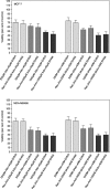
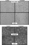
In this study we have examined the cytotoxic effects of different concentrations of adenosine (Ado) and deoxyadenosine (dAdo) on human breast cancer cell lines. Ado and dAdo alone had little effect on cell cytotoxicity. However, in the presence of adenosine deaminase (ADA) inhibitor, EHNA, adenosine and deoxyadenosine led to significant growth inhibition of cells of the lines tested. Ado/EHNA and dAdo/EHNA-induced cell death was significantly inhibited by NBTI, an inhibitor of nucleoside transport, and 5'-amino-5'-deoxyadenosine, an inhibitor of adenosine kinase, but the effects were not affected by 8-phenyltheophylline, a broad inhibitor of adenosine receptors. The Ado/EHNA combination brought about morphological changes consistent with apoptosis. Caspase-9 activation was observed in MCF-7 and MDA-MB468 human breast cancer cell lines on treatment with Ado/EHNA or dAdo/EHNA, but, as expected, caspase-3 activation was only observed in MDA-MB468 cells. The results of the study, thus, suggest that extracellular adenosine and deoxyadenosine induce apoptosis in both oestrogen receptor-positive (MCF-7) and also oestrogen receptor-negative (MDA-MB468) human breast cancer cells by its uptake into the cells and conversion to AMP (dAMP) followed by activation of nucleoside kinase, and finally by the activation of the mitochondrial/intrinsic apoptotic pathway.
Figures


Similar articles
-
Adenosine-mediated killing of cultured epithelial cancer cells.Cancer Res. 2000 Apr 1;60(7):1887-94. Cancer Res. 2000. PMID: 10766176
-
8-Amino-adenosine activates p53-independent cell death of metastatic breast cancers.Mol Cancer Ther. 2012 Nov;11(11):2495-504. doi: 10.1158/1535-7163.MCT-12-0085. Epub 2012 Sep 12. Mol Cancer Ther. 2012. PMID: 22973058 Free PMC article.
-
2'-deoxyadenosine induces apoptosis in rat chromaffin cells.J Neurochem. 1996 Dec;67(6):2273-81. doi: 10.1046/j.1471-4159.1996.67062273.x. J Neurochem. 1996. PMID: 8931458
-
Inhibition of macrophage phagocytosis by methylation inhibitors. Lack of correlation of protein carboxymethylation and phospholipid methylation with phagocytosis.J Biol Chem. 1985 Jan 10;260(1):546-54. J Biol Chem. 1985. PMID: 3871198
-
Effects of adenosine deaminase inhibitors on lymphocyte-mediated cytolysis.Ann N Y Acad Sci. 1985;451:215-26. doi: 10.1111/j.1749-6632.1985.tb27112.x. Ann N Y Acad Sci. 1985. PMID: 3878118
Cited by
-
Adenosine protects against suicidal erythrocyte death.Pflugers Arch. 2007 Jun;454(3):427-39. doi: 10.1007/s00424-007-0218-2. Epub 2007 Feb 7. Pflugers Arch. 2007. PMID: 17285297
-
Thymol protects against 6-hydroxydopamine-induced neurotoxicity in in vivo and in vitro model of Parkinson's disease via inhibiting oxidative stress.BMC Complement Med Ther. 2022 Feb 10;22(1):40. doi: 10.1186/s12906-022-03524-1. BMC Complement Med Ther. 2022. PMID: 35144603 Free PMC article.
-
Adenosine induces cell cycle arrest and apoptosis via cyclinD1/Cdk4 and Bcl-2/Bax pathways in human ovarian cancer cell line OVCAR-3.Tumour Biol. 2013 Apr;34(2):1085-95. doi: 10.1007/s13277-013-0650-1. Epub 2013 Jan 24. Tumour Biol. 2013. PMID: 23345014
-
Discovery and antitumor activities of constituents from Cyrtomium fortumei (J.) Smith rhizomes.Chem Cent J. 2013 Feb 4;7(1):24. doi: 10.1186/1752-153X-7-24. Chem Cent J. 2013. PMID: 23379693 Free PMC article.
-
Metabolomics and metagenomics reveal the impact of γδ T inhibition on gut microbiota and metabolism in periodontitis-promoting OSCC.mSystems. 2024 Feb 20;9(2):e0077723. doi: 10.1128/msystems.00777-23. Epub 2024 Jan 23. mSystems. 2024. PMID: 38259106 Free PMC article.
References
-
- Abbracchio MP, Ceruti S, Brambilla R, Franceschi C, Malorni W, Jacobson KA, von Lubitz DK, Cattabeni F (1997) Modulation of apoptosis by adenosine in the central nervous system: a possible role for the A3 receptor. Pathophysiological significance and therapeutic implications for neurodegenerative disorders. Ann. NY Acad. Sci. 825, 11. - PMC - PubMed
-
- Barczyk K, Kreuter M, Pryjma J, Booy EP, Maddika S, Ghavami S, Berdel WE, Roth J, Los M (2005) Serum cytochrome c indicates in vivo apoptosis and it can serve as a prognostic marker during cancer therapy. Int. J. Cancer 114, 167. - PubMed
-
- Barry CP, Lind SE (2000) Adenosine‐mediated killing of cultured epithelial cancer cells. Cancer Res. 60, 1887. - PubMed
-
- Bradford MM (1976) A rapid and sensitive method for the quantities of microgram of protein utilizing the principle of protein‐dye binding. Anal. Biochem. 72, 248. - PubMed
Publication types
MeSH terms
Substances
LinkOut - more resources
Full Text Sources
Other Literature Sources
Medical
Research Materials

