. 2005 Dec 2;280(48):39914-24.
doi: 10.1074/jbc.M502820200. Epub 2005 Oct 3.

The CATERPILLER protein monarch-1 is an antagonist of toll-like receptor-, tumor necrosis factor alpha-, and Mycobacterium tuberculosis-induced pro-inflammatory signals

Affiliations

The CATERPILLER protein monarch-1 is an antagonist of toll-like receptor-, tumor necrosis factor alpha-, and Mycobacterium tuberculosis-induced pro-inflammatory signals

Kristi L Williams et al. J Biol Chem. .

Abstract

The CATERPILLER (CLR, also NOD and NLR) proteins share structural similarities with the nucleotide binding domain (NBD)-leucine-rich repeat (LRR) superfamily of plant disease-resistance (R) proteins and are emerging as important immune regulators in animals. CLR proteins contain NBD-LRR motifs and are linked to a limited number of distinct N-terminal domains including transactivation, CARD (caspase activation and recruitment), and pyrin domains (PyD). The CLR gene, Monarch-1/Pypaf7, is expressed by resting primary myeloid/monocytic cells, and its expression in these cells is reduced by Toll-like receptor (TLR) agonists tumor necrosis factor (TNF) alpha and Mycobacterium tuberculosis. Monarch-1 reduces NFkappaB activation by TLR-signaling molecules MyD88, IRAK-1 (type I interleukin-1 receptor-associated protein kinase), and TRAF6 (TNF receptor (TNFR)-associated factor) as well as TNFR signaling molecules TRAF2 and RIP1 but not the downstream NFkappaB subunit p65. This indicates that Monarch-1 is a negative regulator of both TLR and TNFR pathways. Reducing Monarch-1 expression with small interference RNA in myeloid/monocytic cells caused a dramatic increase in NFkappaB activation and cytokine expression in response to TLR2/TLR4 agonists, TNFalpha, or M. tuberculosis infection, suggesting that Monarch-1 is a negative regulator of inflammation. Because Monarch-1 is the first CLR protein that interferes with both TLR2 and TLR4 activation, the mechanism of this interference is significant. We find that Monarch-1 associates with IRAK-1 but not MyD88, resulting in the blockage of IRAK-1 hyperphosphorylation. Mutants containing the NBD-LRR or PyD-NBD also blocked IRAK-1 activation. This is the first example of a CLR protein that antagonizes inflammatory responses initiated by TLR agonists via interference with IRAK-1 activation.


Figures
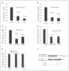
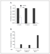
FIGURE 1
FIGURE 1. TLR activation reduces Monarch-1 expression
A, Monarch-1 expression in human adherent peripheral blood cells stimulated for 1 h with commercial LPS (TLR4 agonist) or commercial peptidoglycan (PGN) (TLR2 agonist). Seven separate cell preparations were used. B, Monarch-1 expression in human peripheral blood granulocytes stimulated with commercial LPS (TLR4 agonist) or commercial peptidoglycan (TLR2 agonist). Three separate cell preparations were used. C, Monarch-1 expression in human peripheral blood granulocytes stimulated with purified phenol LPS (TLR4 agonist) or synthetic Pam3Cys (TLR2 agonist). Two separate cell preparations were used. D, Monarch-1 expression in human adherent peripheral blood cells stimulated with purified phenol LPS (TLR4 agonist) or TNFα for 1 h (black bars) and 6 h (white bars). Data are representative of two separate experiments. E, Monarch-1 expression in the human THP-1 monocyte cell line stimulated with phenol-purified LPS for the indicated time points. Data are the average of three separate experiments. Monarch-1 expression was normalized to the expression of 18 S rRNA and are represented as -fold difference compared with control. Error bars represent the S.E. of separate experiments. F, Monarch-1 protein expression in the human THP-1 monocyte cell line stimulated with Pam3Cys for the indicated time points. Lysates were immuno-precipitated (IP) with rabbit anti-Monarch-1 and rat anti-actin followed by immunoblotting (IB) with either mouse anti-Monarch-1 or goat anti-actin-HRP antibodies. Data are representative of two separate experiments.
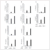
FIGURE 2
FIGURE 2. Monarch-1 blocks signaling molecules in the TLR and TNFR pathways
A, HEK293T cells were co-transected with TLR4, CD14, MD-2, different amounts of Monarch-1, and an ELAM-luciferase reporter construct. After 24 h cells were stimulated with the indicated concentration of purified E. coli K235 LPS for 5 h. Results represent the mean ± S.E. of three separate experiments. B, HEK293T cells were co-transfected with either vector alone or expression plasmids for MyD88, IRAK-1, TRAF6, or P65 with/without three concentrations of Monarch-1, and an NFκB luciferase reporter construct. The results represent the mean ± S.E. of three separate experiments. C, HEK293T cells were co-transfected with either vector alone or expression plasmids for TRAF2 or RIP1 or stimulated for 8 h with TNFαwith/without three concentrations of Monarch-1 and an NFκB luciferase reporter construct. As a control, cells were co-transfected with an expression plasmid for class II transactivator (CIITA) and an HLA-DR luciferase reporter construct plus either vector alone or the highest concentration of Monarch-1 plasmid. The results are representative of four separate experiments.
FIGURE 3
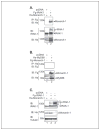
FIGURE 3. Monarch-1 associates with IRAK-1 and limits the accumulation of hyperphosphorylated forms of the kinase
A, HEK293T cells were transfected with the indicated plasmid DNA. Twenty-four hours later whole cell lysates (WCL) were prepared, and FLAG-IRAK-1-containing protein complexes were immunoprecipitated (IP) with anti-FLAG (Fg) M2-agarose beads and separated by SDS-PAGE. Western blots (IB) were probed with anti-HA (Ha) antibody to detect co-precipitating HA-Monarch-1. p-, phosphorylated. B, cells were transfected with empty vector or expression vectors for FLAG-tagged Monarch-1 and HA-MyD88. MyD88-containing protein complexes were immunoprecipitated with an anti-HA antibody and separated by SDS-PAGE. Western blots were probed with anti-FLAG to detect co-precipitating FLAG-Monarch-1. C, cells were transfected with the indicated plasmid DNA, and 24 h later whole cell lysates were prepared and separated by SDS-PAGE. Western blots were probed with the indicated antibodies. The hyperphosphorylated form of IRAK-1 (marked as III) was greatly reduced by the presence of Monarch-1.
FIGURE 4
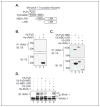
FIGURE 4. The Monarch-1 NBD is required for the association and regulation of IRAK-1 hyperphosphorylation
A, diagram depicting V5-tagged Monarch-1 truncation mutants. B, cells were transfected with expression vectors encoding the V5-tagged PyD of Monarch-1 and HA-IRAK-1. IRAK-1-containing protein complexes were immunoprecipitated (IP) with an anti-IRAK-1 antibody and separated by SDS-PAGE. Western blots (IB) were probed with anti-V5 to detect co-precipitating V5-PyD. C, cells were transfected with expression vectors encoding the indicated V5-tagged truncation mutant of Monarch-1 in addition to HA-IRAK-1. IRAK-1-containing protein complexes were immunoprecipitated with an anti-IRAK-1 antibody and separated by SDS-PAGE. Western blots were probed with anti-V5 to detect co-precipitating V5-tagged Monarch-1 truncation mutants. D, whole cell lysates derived from experiments described in A and B were separated by SDS-PAGE and probed with an anti-IRAK-1 antibody. The hyperphosphorylated form of IRAK-1 (marked as III) was greatly reduced in the presence of the NBD-LRR-containing truncation mutant of Monarch-1.
FIGURE 5
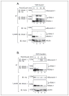
FIGURE 5. Monarch-1 associates with endogenous IRAK-1 in THP-1 monocytic cells upon TLR stimulation and limits the accumulation of hyperphosphorylated, endogenous IRAK-1
THP-1 cells stably expressing (A) empty vector (THP-HA-EV) or (B) HA-tagged Monarch-1 (THP-HA-Mon) were stimulated with the TLR2 agonist Pam3Cys4 for the indicated times. Endogenous IRAK-1-containing protein complexes were immunoprecipitated (IP) with an anti-IRAK-1 antibody and then fractionated by SDS-PAGE. Western blots were probed with anti-HA to detect co-precipitating HA-Monarch-1 (top panels). These immunoblots (IB) were then stripped and probed with anti-IRAK-1 antibodies (second panels). The hyperphosphorylated form of IRAK-1 was greatly reduced by the presence of Monarch-1. Interestingly, analysis of cellular lysates indicated that the disappearance of the 80-kDa nonphosphorylated form of IRAK-1 occurs more slowly in the presence of Monarch-1. Actin immunoblots were performed to ensure equivalent levels of protein were used among the samples. p-, phosphorylated.
FIGURE 6
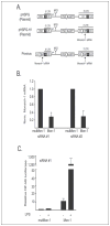
FIGURE 6. Monarch-1 is a negative regulator of NFκB activity in the THP-1 monocytic cell line
A, plasmid map of Monarch-1 siRNA in the pHSPG retroviral vector. SiRNAs specific for Monarch-1 were developed to block Monarch-1 expression followed by analysis of gene activity. Monarch-1 siRNAs, driven by the H1 RNA promoter, were inserted into the pHSPG retroviral vector within the 3′ LTR. The presence of a phosphoglycerate kinase (PGK) promoter driving enhanced green fluorescent protein (eGFP) allows for selection of transduced cells by fluorescence-activated cell sorting. Two separate Monarch-1 siRNAs and their corresponding mutant, controls were placed in pHSPG vectors. B, analysis of Monarch-1 expression in Monarch-1 (Mon-1) and control mutant (mutMon-1) siRNA bulk monocyte THP-1 cultures as determined by real-time PCR. THP-1 cells were infected with two different Monarch-1-specific siRNAs, siRNA#1 and siRNA#2, respectively. Expression was normalized to the expression of 18 S mRNA and are represented as -fold over mutant control. The results represent the mean ± S.E. of five separate experiments. C, analysis of NFκB activity in Monarch-1-specific siRNA cultures. Monarch-1 (Mon-1) and control mutant (mutMon-1) siRNA cultures were infected with a retroviral based NFκB luciferase reporter. Cultures were then stimulated with E. coli K235 LPS followed by analysis of NFκB luciferase activity. Expression is represented as -fold over mutant control. Results represent the mean ± S.E. of three separate experiments.
FIGURE 7
FIGURE 7. Monarch-1 is a negative regulator of IL-6 in monocytic THP-1 cells induced by LPS
Analysis of IL-6 mRNA and protein expression in Monarch-1-specific siRNA cultures. A, two different Monarch-1 (Mon-1) and control mutant (mut-Mon-1) siRNA cultures, siRNA#1 and siRNA#2, were stimulated with E. coli K235 LPS followed by analysis of IL-6 transcript expression by real-time PCR. IL-6 expression was normalized to 18 S RNA and are represented as exponential numbers. Data are a representative of five separate experiments. B, two different Monarch-1 (Mon-1) and control mutant (mutMon-1) siRNA cultures, siRNA#1 and siRNA#2, were stimulated with E. coli K235 LPS followed by analysis of IL-6 cytokine expression by ELISA. Data are representative of three separate experiments. ND, not determined.
FIGURE 8
FIGURE 8. Monarch-1 is a negative regulator of IL-6 in the THP-1 monocytic cell line infected with virulent M. tuberculosis
A, Monarch-1 transcript expression in the human THP-1 monocytic cell line infected with virulent M. tuberculosis (Mtb) for the indicated time points. Data represent the average of three separate experiments measured by real-time PCR. B, analysis of IL-6 mRNA expression in Monarch-1-specific siRNA cultures. THP-1 cells were compared with Monarch-1 (Mon-1) and control mutant (mut-Mon-1) siRNA cultures after infection with M. tuberculosis followed by analysis of IL-6 expression by real-time PCR. Expression was normalized to 18 S RNA, and data are shown as -fold over unstimulated control. Data are representative of three separate experiments.
FIGURE 9
FIGURE 9. Monarch-1 is a negative regulator of cytokine induction by TLR4 and TLR2 agonists as well as the TNFR pathway
Monarch-1 (Mon-1) and control mutant (mutMon-1) siRNA cultures were stimulated for 24 h with Pam3Cys (TLR2 agonist) (A), E. coli K235 LPS (TLR4 agonist) (B), or TNFα (C) followed by analysis of cytokine expression by cytokine bead assay. Results represent the mean ± S.E. of three separate experiments.

References

    1. Harton JA, Linhoff MW, Zhang J, Ting JP. J Immunol. 2002;169:4088–4093. - PubMed
    1. Inohara N, Nunez G. Nat Rev Immunol. 2003;3:371–382. - PubMed
    1. Tschopp J, Martinon F, Burns K. Nat Rev Mol Cell Biol. 2003;4:95–104. - PubMed
    1. Pawlowski K, Pio F, Chu Z, Reed JC, Godzik A. Trends Biochem Sci. 2001;26:85–87. - PubMed
    1. Martinon F, Hofmann K, Tschopp J. Curr Biol. 2001;11:118–120. - PubMed

Publication types

MeSH terms

Substances