. 2005 Oct 10;171(1):27-33.
doi: 10.1083/jcb.200504107. Epub 2005 Oct 3.

Rootletin forms centriole-associated filaments and functions in centrosome cohesion

Affiliations

Rootletin forms centriole-associated filaments and functions in centrosome cohesion

Susanne Bahe et al. J Cell Biol. .

Abstract

After duplication of the centriole pair during S phase, the centrosome functions as a single microtubule-organizing center until the onset of mitosis, when the duplicated centrosomes separate for bipolar spindle formation. The mechanisms regulating centrosome cohesion and separation during the cell cycle are not well understood. In this study, we analyze the protein rootletin as a candidate centrosome linker component. As shown by immunoelectron microscopy, endogenous rootletin forms striking fibers emanating from the proximal ends of centrioles. Moreover, rootletin interacts with C-Nap1, a protein previously implicated in centrosome cohesion. Similar to C-Nap1, rootletin is phosphorylated by Nek2 kinase and is displaced from centrosomes at the onset of mitosis. Whereas the overexpression of rootletin results in the formation of extensive fibers, small interfering RNA-mediated depletion of either rootletin or C-Nap1 causes centrosome splitting, suggesting that both proteins contribute to maintaining centrosome cohesion. The ability of rootletin to form centriole-associated fibers suggests a dynamic model for centrosome cohesion based on entangling filaments rather than continuous polymeric linkers.


Figures

Figure 1.
Figure 1.
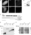
Human rootletin localizes to centrosomes. (A) U2OS cells were costained with antirootletin (green) and anti–γ-tubulin (red) antibodies. (a) Interphase cells; insets show enlargements of centrosomes. (b–d) Mitotic cells showing prophase (b), metaphase (c), and telophase (d). DNA was stained with DAPI. Bars, 5 μm. (B) Western blots. (left) Antirootletin antibody R145 on total extracts from myc-rootletin–transfected (lane 1) or untransfected (lane 2) 293T cells. (right) Preimmune (P, lane 3) and antirootletin (I, lane 4) serum on centrosomes isolated from KE37 cells. Arrowheads point to human rootletin. (C) U2OS cells were subjected to preembedding immunogold-labeling EM. Cells were labeled with either antirootletin antibody R145 followed by Nanogold-coupled secondary antibody (a and c), secondary antibody alone (b), or antibodies directed against the NH2 terminus of rootletin (R146; d). Brackets (d) emphasize the distance of gold particles from the centriole surface with R146 labeling. Bars, 250 nm.
Figure 2.
Figure 2.
Rootletin interacts with itself, C-Nap1, and Nek2. (A) U2OS cells were transfected with myc-tagged rootletin (green) and were counterstained with anti–γ-tubulin antibody (red). Insets show enlargements of the centrosomes to highlight protein fibers. Bar, 5 μm. (B) U2OS cells were transfected with tagged Nek2, C-Nap1, or a COOH-terminal fragment of C-Nap1 either alone (right) or together with rootletin (left three panels), and the distribution of proteins was monitored by IF microscopy. Bars, 15 μm. (C) Yeast two-hybrid interaction of rootletin with Nek2 (top) and C-Nap1 (bottom). Transformed yeast cells were plated onto media selecting for transformants (−LW) and bait-prey interactions (−QDO), respectively. AD, activation domain; BD, binding domain; LW, leucine tryptophane; QDO, quadruple drop out.
Figure 3.
Figure 3.
Nek2 phosphorylates rootletin. (A) U2OS cells were transfected with wild-type (wt) or catalytically inactive (K37R) GFP-Nek2, and endogenous rootletin was stained with antibody. Bars, 5 μm. (B–D) After the coexpression of GFP-Nek2 with full-length myc-rootletin (B) or myc-rootletin fragments (D, arrowheads), total 293T cell extracts were probed by Western blotting with anti-myc antibody. (C) Schematic illustrating rootletin fragments (black bars indicate predicted coiled-coil). (E) In vitro kinase assay using wild-type or K37R Nek2 on His-rootletin NH2- and COOH-terminal fragments. Proteins were resolved by SDS-PAGE, and gels were subjected to autoradiography (left) and Coomassie blue staining (right). Arrowheads indicate the migration of His-rootletin fragments; the arrow points to autophosphorylated Nek2.
Figure 4.
Figure 4.
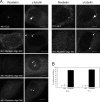
siRNA depletion of rootletin causes centrosome splitting. (A) U2OS cells were transfected for 48 or 72 h with control (GL2) or different rootletin-specific siRNA duplexes and were costained for rootletin and γ-tubulin. Arrows and arrowheads mark paired centrioles and split centrosomes, respectively. Bars, 10 μm. (B) Quantitation of centrosome splitting in control (GL2) or rootletin siRNA-treated cells (oligonucleotide 222). Centrosomes were counted as split when the distance between centrioles was >2 μm. Error bars represent SEM.
Figure 5.
Figure 5.
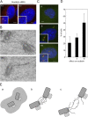
Mutual dependency of rootletin and C-Nap1 localization and models for centrosome cohesion. (A) U2OS cells were transfected for 48 or 72 h with control (GL2) or rootletin-specific siRNA duplexes and were stained for C-Nap1 (green) and γ-tubulin (red). Bars, 5 μm. (B) U2OS cells were depleted of C-Nap1 by siRNA and were subjected to immuno-EM labeling of rootletin (antibody R145). Bars, 250 nm. (C) U2OS cells were transfected for 48 h with a C-Nap1–specific siRNA duplex and were stained for rootletin (red) and γ-tubulin (green). Rootletin disappeared altogether (I) and formed elongated fibers that either appeared to connect split centrosomes (II) or protruded away from split centrosomes (III). Bars, 5 μm. (A and C) Insets show enlargements of the centrosome area to highlight centrosome splitting. (D) Quantitation of the observed effects of C-Nap1 RNA interference on rootletin localization (I–III; compare with C). Error bars represent SEM. (E) Schematic illustration of possible models for centrosome cohesion. (a) Centrioles (rectangles) embedded in the PCM (speckled). (b) Centrioles connected by a continuous proteinaceous linker. (c) Centrioles connected by entangling filaments. C, C-Nap1; L, hypothetical continuous linker; R, rootletin; X, additional linker proteins that are yet to be identified. Parental centrioles (dark gray) are depicted as having associated nascent procentrioles (dotted lines).

References

    1. Andersen, J.S., C.J. Wilkinson, T. Mayor, P. Mortensen, E.A. Nigg, and M. Mann. 2003. Proteomic characterization of the human centrosome by protein correlation profiling. Nature. 426:570–574. - PubMed
    1. Bornens, M. 2002. Centrosome composition and microtubule anchoring mechanisms. Curr. Opin. Cell Biol. 14:25–34. - PubMed
    1. Bornens, M., M. Paintrand, J. Berges, M.C. Marty, and E. Karsenti. 1987. Structural and chemical characterization of isolated centrosomes. Cell Motil. Cytoskeleton. 8:238–249. - PubMed
    1. Doxsey, S., D. McCollum, and W. Theurkauf. 2005. Centrosomes in cellular regulation. Annu. Rev. Cell Dev. Biol 10.1146/annurev.cellbio. 21.122303.120418. - DOI - PubMed
    1. Elbashir, S.M., J. Harborth, W. Lendeckel, A. Yalcin, K. Weber, and T. Tuschl. 2001. Duplexes of 21-nucleotide RNAs mediate RNA interference in cultured mammalian cells. Nature. 411:494–498. - PubMed

Publication types

MeSH terms