Comparative Study
. 2005 Oct 3;202(7):987-99.
doi: 10.1084/jem.20051239.

The human macrophage mannose receptor directs Mycobacterium tuberculosis lipoarabinomannan-mediated phagosome biogenesis

Affiliations
Comparative Study

The human macrophage mannose receptor directs Mycobacterium tuberculosis lipoarabinomannan-mediated phagosome biogenesis

Peter B Kang et al. J Exp Med. .

Abstract

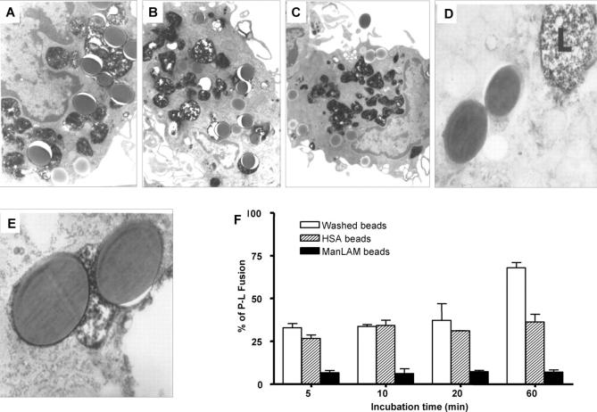
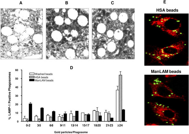
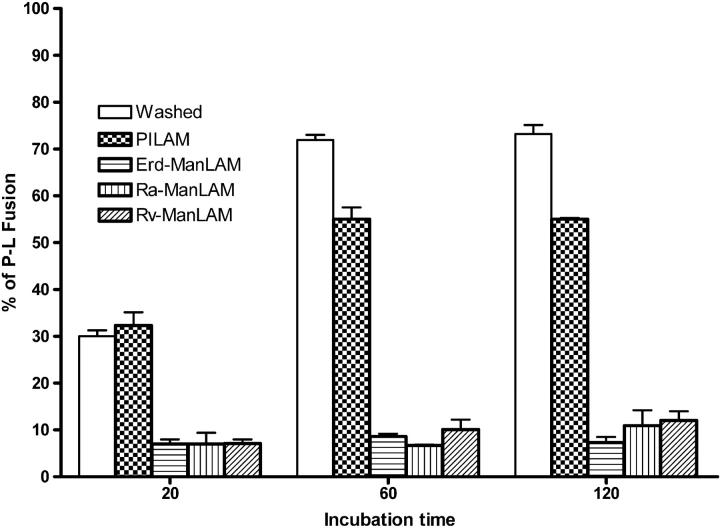
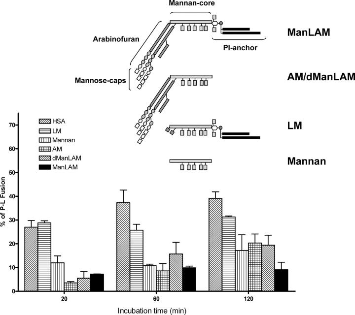
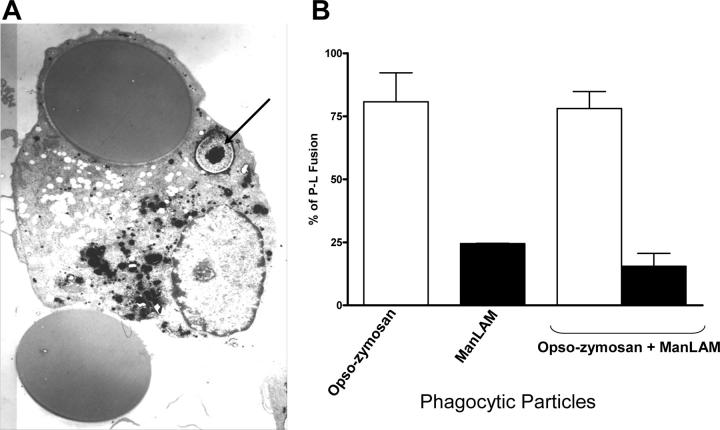
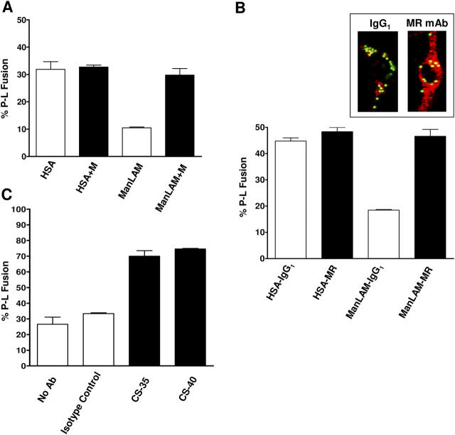
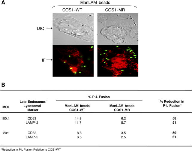
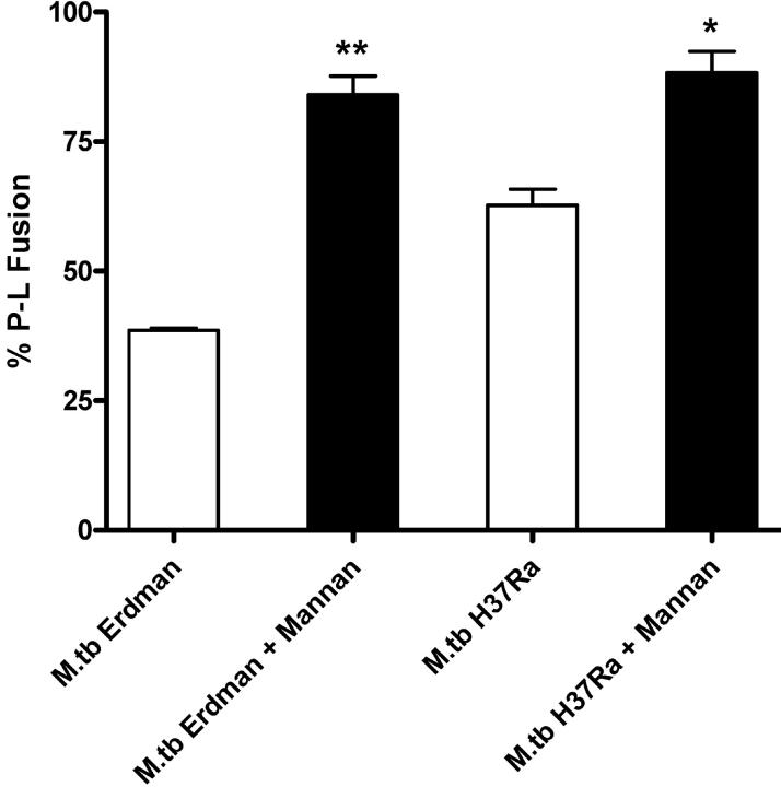
Mycobacterium tuberculosis (M.tb) survives in macrophages in part by limiting phagosome-lysosome (P-L) fusion. M.tb mannose-capped lipoarabinomannan (ManLAM) blocks phagosome maturation. The pattern recognition mannose receptor (MR) binds to the ManLAM mannose caps and mediates phagocytosis of bacilli by human macrophages. Using quantitative electron and confocal microscopy, we report that engagement of the MR by ManLAM during the phagocytic process is a key step in limiting P-L fusion. P-L fusion of ManLAM microspheres was significantly reduced in human macrophages and an MR-expressing cell line but not in monocytes that lack the receptor. Moreover, reversal of P-L fusion inhibition occurred with MR blockade. Inhibition of P-L fusion did not occur with entry via Fcgamma receptors or dendritic cell-specific intracellular adhesion molecule 3 grabbing nonintegrin, or with phosphatidylinositol-capped lipoarabinomannan. The ManLAM mannose cap structures were necessary in limiting P-L fusion, and the intact molecule was required to maintain this phenotype. Finally, MR blockade during phagocytosis of virulent M.tb led to a reversal of P-L fusion inhibition in human macrophages (84.0 +/- 5.1% vs. 38.6 +/- 0.6%). Thus, engagement of the MR by ManLAM during the phagocytic process directs M.tb to its initial phagosomal niche, thereby enhancing survival in human macrophages.


Figures

Figure 1.
Figure 1.
ManLAM bead phagosomes demonstrate limited fusion with lysosomes in macrophages after phagocytosis. Low-power electron micrographs of P-L fusion events using (A) washed beads (×24,000), (B) HSA beads (×14,000) and (C) ManLAM beads (×12,000). Lysosomes contain black particulate material. High-power electron micrographs of (D) ManLAM bead phagosomes shown with a neighboring lysosome (L) that is not fused (×40,000), and of (E) washed beads fused with lysosomes forming a phagolysosome (×60,000). (F) Extent of P-L fusion of washed, HSA, and ManLAM bead phagosomes. MDMs in monolayer culture were pulsed/chased with 1 mg/ml horseradish peroxidase for 2 h at 37°C. A synchronized phagocytosis assay was performed for the indicated times. After fixation, MDMs were processed for TEM. The percentage of P-L fusion (mean ± SEM, n = 2) was significantly reduced with ManLAM beads compared with both washed and HSA bead controls (P < 0.005; one-way ANOVA).
Figure 2.
Figure 2.
LAMP-1 staining on ManLAM bead phagosomes in macrophages is reduced. Electron micrographs (×20,000) of (A) washed and (B) HSA bead phagosomes show higher levels of LAMP-1 accumulation than (C) ManLAM bead phagosomes. (D) A synchronized phagocytosis assay was performed for 60 min. After fixation, MDMs were processed for immunohistochemical TEM. The distribution of LAMP-1 staining is shown as the percentage of LAMP-1–positive phagosomes containing washed, HSA, or ManLAM beads (mean ± SEM, n = 2). (E) HSA bead phagosomes show a higher degree of LAMP-1 colocalization than ManLAM bead phagosomes in macrophages following phagocytosis. MDM monolayers were incubated with either HSA or ManLAM fluorescent beads for 2 h. After washing, monolayers were fixed, permeabilized, and incubated with mouse anti–human LAMP-1 mAb followed by goat anti–mouse IgG secondary antibody. LAMP-1 colocalization was enumerated by confocal microscopy. Compartments that stain positive for LAMP-1 are red, beads in unfused phagosomes are green, and beads in fused phagolysosomes are yellow. The photomicrographs are from one representative experiment (n = 3).
Figure 3.
Figure 3.
ManLAM from M.tb strains but not PILAM from M. smegmatis limits P-L fusion in macrophages. Washed, PILAM-, Ra-ManLAM-, Rv-ManLAM-, and Erd-ManLAM-coated beads were incubated with MDMs for the indicated times in the synchronized phagocytosis assay. The extent of P-L fusion of bead phagosomes was examined and enumerated via TEM as described in Fig. 1 F (mean ± SEM, n = 2).
Figure 4.
Figure 4.
The terminal mannosyl caps of ManLAM are important in limiting P-L fusion. Beads coated with HSA, intact ManLAM or its substructures LM, mannan, arabinomannan, and dManLAM were used with MDMs in the synchronized phagocytosis assay for the indicated times. P-L fusion was examined and enumerated via TEM as in Fig. 1 F (mean ± SEM, n = 2).
Figure 5.
Figure 5.
Limited P-L fusion is restricted to the ManLAM bead phagosome. MDM monolayers were incubated with ManLAM beads (20 μm) for 1 h followed by the addition of opsonized zymosan for 1 h before fixation. P-L fusion was analyzed by TEM as in Fig. 1. (A) Electron micrograph (×10,000) shows one unfused ManLAM bead phagosome along with an opsonized zymosan particle in a phagolysosome (arrow). A second ManLAM miscrosphere (shown at the bottom surface of the cell) is forming a phagocytic cup on the cell membrane. (B) The bar graph shows that the levels of P-L fusion for either opsonized zymosan or ManLAM beads phagocytosed individually by MDMs do not change when both types of particles are phagocytosed by the same cell. Shown is a representative experiment (n = 3).
Figure 6.
Figure 6.
MR blockade with mannan or anti-MR antibody and redirected entry via FcγRs reverse the inhibition of P-L fusion of ManLAM beads in macrophages to control levels. (A) MDMs were preincubated with or without mannan for 30 min before adding HSA or ManLAM beads and allowing for synchronized phagocytosis over 60 min. P-L fusion was examined and enumerated by TEM as in Fig. 1 F (mean ± SEM, n = 2) and was significantly reduced with untreated ManLAM beads compared with the HSA control and mannan-treated bead groups (P < 0.005, one-way ANOVA). (B) MDMs were preincubated with anti-MR or control IgG1 mAbs (10 μg/ml) for 20 min at 37°C followed by the addition of either HSA or ManLAM beads (MOI 100–200:1), and phagocytosis assay was then performed for 2 h. The cells were fixed, permeabilized, blocked, and then stained with anti-CD63 mAb followed by Alexa Fluor 647-conjugated mouse IgG. P-L fusion was examined and enumerated by confocal microscopy (mean ± SEM, n = 2). Data were analyzed via one-way ANOVA comparing the value of the ManLAM bead group (IgG1 mAb-treated MDMs) with those of the HSA bead control groups and the ManLAM bead group (MR mAb-treated MDMs) (P < 0.001). Inset: A control IgG1 mAb-treated MDM shows limited P-L fusion (left panel) and an anti-MR mAb-treated MDM shows increased P-L fusion (right panel) of ManLAM bead phagosomes. CD63-positive compartments are red, beads in unfused phagosomes are green, and those colocalized with CD63 are yellow. (C) ManLAM beads were preincubated with anti-LAM mAbs CS-35 or CS-40, IgG subtype control, or no mAb at 37°C for 60 min, washed, and added to MDM monolayers for the synchronized phagocytosis assay (60 min). P-L fusion was examined and quantified via TEM as in Fig. 1 F (mean ± SEM, n =2) and was significantly increased in the groups treated with either CS-35 or CS-40 compared with each control group (P < 0.0005, one-way ANOVA).
Figure 7.
Figure 7.
ManLAM beads demonstrate reduced P-L fusion in MR-expressing COS-1 cells. COS1-WT and COS1-MR cells adhered to glass coverslips were incubated with ManLAM beads (MOI 20:1 or 100:1) for 3 h using a synchronized phagocytosis assay. Cell monolayers were fixed, permeabilized, and stained with anti–human CD63 or LAMP-2 mAbs followed by Alexa Fluor 647-conjugated goat anti–mouse IgG. P-L fusion was examined and enumerated via confocal microscopy. (A) Phase and fluorescence microscopy images of ManLAM beads in COS1-WT and COS1-MR cells. CD63 positive compartments are red, beads in unfused phagosomes are green, and those colocalized with CD63 are yellow. Shown is a representative experiment (n = 2). DIC, differential interference contrast; IF, immunofluorescence. (B) Percent P-L fusion of ManLAM beads using two different MOIs (100:1 and 20:1, n = 2, performed in duplicate).
Figure 8.
Figure 8.
MR blockade increases the level of P-L fusion of phagosomes containing live M.tb bacilli following phagocytosis. MDMs were preincubated with or without mannan at 37°C for 30 min followed by incubation with the live M.tb strains Erdman and H37Ra (MOI of 6.25 bacilli per MDM) for 2 h. P-L fusion was examined and quantified via TEM as in Figure 1 F (mean ± SEM, n = 2). **P < 0.01 (Erdman) and *P < 0.05 (H37Ra) relative to the no mannan condition for each strain.
Figure 9.
Figure 9.
Heat-killing and γ-irradiation of M.tb alter the integrity of the bacterial cell wall and decrease the amount of surface-exposed ManLAM. Single suspensions of plate-grown M.tb strain Erdman were kept alive or were killed by heating or γ-irradiation. Bacilli were fixed with 2% paraformaldehyde, washed, embedded in LR White, thin sectioned, and immunostained with anti-LAM mAb CS-35 or CS-40 followed by 10 nm immunogold-conjugated goat anti–mouse secondary antibody before viewing by TEM. Electron micrographs of (A) live M.tb stained with CS-35 (bar, 100 nm), (B) live M.tb stained with CS-40 (bar, 100 nm), or (C) heat-killed M.tb stained with CS-35 (bar, 100 nm). (D) Graph shows percentage of bacilli in the indicated groups that have an intact cell wall architecture. (E) Graph shows amount of ManLAM exposed on the surface of live, heat-killed, and γ-irradiated M.tb. Results are shown as a percentage of immunogold particles/M.tb bacillus relative to control live M.tb set at 100% (*P < 0.05).

References

    1. Fenton, M.J., L.W. Riley, and L.S. Schlesinger. 2005. Receptor-mediated recognition of Mycobacterium tuberculosis by host cells. Tuberculosis and the Tubercle Bacillus. S.T. Cole, K.D. Eisenach, D.N. McMurray, and W.R. Jacobs, Jr., editors. ASM Press, New York. 405–426.
    1. Brennan, P.J., and H. Nikaido. 1995. The envelope of mycobacteria. Annu. Rev. Biochem. 64:29–63. - PubMed
    1. Strohmeier, G.R., and M.J. Fenton. 1999. Roles of lipoarabinomannan in the pathogenesis of tuberculosis. Microbes Infect. 1:709–717. - PubMed
    1. Hunter, S.W., and P.J. Brennan. 1990. Evidence for the presence of a phosphatidylinositol anchor on the lipoarabinomannan and lipomannan of Mycobacterium tuberculosis. J. Biol. Chem. 265:9272–9279. - PubMed
    1. Armstrong, J.A., and P.D. Hart. 1971. Response of cultured macrophages to Mycobacterium tuberculosis, with observations on fusion of lysosomes with phagosomes. J. Exp. Med. 134:713–740. - PMC - PubMed

Publication types

MeSH terms