Comparative Study
. 2005 Oct 3;202(7):1001-12.
doi: 10.1084/jem.20051143.

Cytolytic granule polarization and degranulation controlled by different receptors in resting NK cells

Affiliations
Comparative Study

Cytolytic granule polarization and degranulation controlled by different receptors in resting NK cells

Yenan T Bryceson et al. J Exp Med. .

Abstract

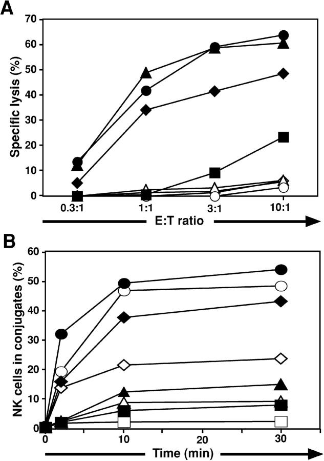
The relative contribution to cytotoxicity of each of the multiple NK cell activation receptors has been difficult to assess. Using Drosophila insect cells, which express ligands of human NK cell receptors, we show that target cell lysis by resting NK cells is controlled by different receptor signals for cytolytic granule polarization and degranulation. Intercellular adhesion molecule (ICAM)-1 on insect cells was sufficient to induce polarization of granules, but not degranulation, in resting NK cells. Conversely, engagement of the Fc receptor CD16 by rabbit IgG on insect cells induced degranulation without specific polarization. Lysis by resting NK cells occurred when polarization and degranulation were induced by the combined presence of ICAM-1 and IgG on insect cells. Engagement of receptor 2B4 by CD48 on insect cells induced weak polarization and no degranulation. However, coengagement of 2B4 and CD16 by their respective ligands resulted in granule polarization and cytotoxicity in the absence of leukocyte functional antigen-1-mediated adhesion to target cells. These data show that cytotoxicity by resting NK cells is controlled tightly by separate or cooperative signals from different receptors for granule polarization and degranulation.


Figures

Figure 1.
Figure 1.
ADCC by resting NK cells toward insect SC2 cells requires costimulation. NK cells were mixed with SC2 (open squares), SC2–ICAM-1 (open diamonds), SC2–CD48 (open triangles), or SC2–ICAM-1–CD48 (open circles) cells or the above SC2 cells were preincubated with a rabbit serum raised against SC2 cells (filled symbols). (A) Cells were incubated for 3 h at 37°C. Specific lysis of SC2 cells was calculated from the percentage of propidium iodide–positive SC2 cells in duplicate samples, as determined by flow cytometry. (B) NK cells labeled with a green dye were mixed with SC2 cells labeled with a red dye, and incubated at 37°C for the indicated time. Cells were fixed and the fraction of NK cells in two-color conjugates was determined by flow cytometry. Points represent the average of duplicate samples. For cytotoxicity, the variation between duplicate samples was, on average, <10% of the calculated value of lysis. Representative experiments are shown.
Figure 2.
Figure 2.
ICAM-1 on insect cells induces granule polarization in resting NK cells. (A) NK cells mixed with SC2 cells expressing indicated ligands were incubated for 20 min at 37°C, fixed, permeabilized, and stained with an antiperforin mAb and fluorochrome-conjugated secondary antibody. NK cells in conjugate with SC2 cells were scored for polarized perforin toward the target cell interface in six independent experiments (three for SC2–ICAM-1–CD48). (B) NK cells were mixed with protein A coupled beads coated with recombinant ICAM-1 Fc. Data are expressed as the fold increase of NK cells that display polarized perforin in three independent experiments. Error bars represent the SD. *P < 0.05 and **P < 0.005 relative to NK cells mixed with SC2 cells or empty beads, respectively.
Figure 3.
Figure 3.
Cell surface CD107a on resting NK cells correlates with granzyme B release and target cell lysis. (A) NK cells were fixed, permeabilized, and stained with anti-CD107a and antiperforin mAbs. Left panels show confocal images of CD107a (green), perforin (red). Right panels show a differential interference contrast (DIC) image, and an overlay of all three images. (B–E) NK cells were mixed with K562 cells or with P815 cells preincubated with the indicated mAbs. (B and D) After 2 h at 37°C, the cells were stained with fluorochrome-conjugated anti-CD56 and anti-CD107a antibodies. NK cells were gated away from target cells on forward scatter/side scatter plots. (B) Profiles show CD56 versus CD107a or control antibody staining, as indicated. (C) In 51Cr release assays, resting NK cells were incubated with K562 cells or with P815 cells incubated with isotype control (open squares), anti-CD16 (filled squares), or anti-2B4 (open triangles) mAbs. Specific lysis obtained in one representative experiment is shown. (D) The fraction of CD107a+ NK cells is presented as the mean of six independent experiments. Bars indicate SD. *P < 0.05 and **P < 0.005 relative to NK cells incubated without target cells. (E) After 2 h, granzyme B release to supernatants was measured by ELISA. Values represent mean ± SD of three independent experiments.
Figure 4.
Figure 4.
CD107a at the surface of degranulated cells. (A and B) Resting NK cells were stained with FITC-conjugated anti-CD56 mAb, fixed, permeabilized, and stained with isotype control (A) or anti-CD107a (B) mAbs. NK cells were gated by forward/side scatter gate. (C and D) Resting NK cells were stained with FITC-conjugated anti-CD56 mAb, fixed, permeabilized, and stained with biotinylated anti-CD107a mAb followed by streptavidin-APC (C) or streptavidin-PE (D). (E–J) Resting NK cells were incubated without (−) or with K562 cells, as indicated on the right. Resting NK cells were incubated for 2 h at 37°C. (F and H) Biotinylated anti-CD107a mAb was included during the incubation. Thereafter, cells were stained with FITC-conjugated anti-CD56 mAb and biotinylated anti-CD107a mAb followed by streptavidin-APC, fixed, permeabilized, and stained with streptavidin-PE (E–H) or biotinylated anti-CD107a mAb followed by streptavidin-PE (I and J). (J) NK cells were treated with cycloheximide. (C–J) Plots are gated on CD56dim NK cells. Numbers indicate the percentage of cells in each quadrant.
Figure 5.
Figure 5.
ICAM on insect cells induce adhesion but not degranulation by resting NK cells. (A) Resting NK cells labeled with a green dye were mixed with SC2 cells labeled with a red dye and incubated at 37°C for 10 min. Cells were fixed and the fraction of NK cells in two-color conjugates was determined by flow cytometry. Values represent the mean of three independent experiments. Bars represent the SD. (B) Resting NK cells were mixed with SC2–ICAM-1 or SC2–ICAM-2, and incubated at 37°C. After 2 h, cells were stained with fluorochrome-conjugated anti-CD56 and anti-CD107a antibodies. NK cells were gated on forward scatter/side scatter plots; the profiles show CD56 versus CD107a antibody staining. The profiles are representative of three or more independent experiments.
Figure 6.
Figure 6.
CD16 engagement induces degranulation in resting NK cells. (A–C) Resting NK cells were mixed with SC2, SC2–ICAM-1, SC2–CD48, or SC2–ICAM-1–CD48. Where indicated, SC2 cells were preincubated with a rabbit anti-SC2 serum (+IgG). Cells were incubated at 37°C for 2 h. (A, B) Thereafter, cells were stained with fluorochrome-conjugated anti-CD107a and anti-CD56 mAbs and analyzed by flow cytometry. (A) NK cells were gated on forward scatter/side scatter plots and profiles show CD56 versus CD107a mAb staining. (B) The percentage of CD56dim CD107a+ NK cells is presented as the mean of six independent experiments. Bars indicate SD. ** P < 0.005 relative to NK cells incubated alone. (C) Granzyme B released in supernatants was measured by ELISA. Values represent mean ± SD of three independent experiments.
Figure 7.
Figure 7.
Rapid coinduction of CD107a and Fas ligand at the surface of resting NK cells. NK cells alone (line), or mixed with SC2 (open squares), SC2–ICAM-1 (open diamonds), SC2–CD48 (open triangles) or SC2–ICAM-1–CD48 (open circles) were incubated for 5, 10, 30, 60, and 120 min at 37°C. For Fc receptor stimulation, SC2 cells also were preincubated with a rabbit anti-SC2 serum (filled symbols). Cells were stained with fluorochrome-conjugated anti-CD56 and anti-CD107a mAbs (A), biotinylated anti-FasL mAb followed by fluorochrome-conjugated streptavidin (B), or fluorochrome-conjugated anti-CD69 mAb (C). The percentage of CD56dimCD107a+ NK cells (A), and the mean fluorescence intensity (MFI) of CD56dim NK cells for Fas ligand (B) and CD69 (C) is shown. Values represent the average of duplicate samples. A representative experiment is shown.
Figure 8.
Figure 8.
2B4-mediated signals lower the threshold for CD16-induced degranulation and increase the number of resting NK cells that degranulate. NK cells were mixed with SC2 (open squares) or SC2–CD48 (open triangles) cells that had been preincubated with serial dilutions of a rabbit anti-SC2 serum. Cells were incubated at 37°C in the presence of no (empty), isotype control (gray shade), or anti-2B4 (filled) mAbs. After 2 h, cells were stained with fluorochrome conjugated anti-CD56 and anti-CD107a mAbs. The mean percentage of CD56dimCD107a+ NK cells of triplicate samples from one representative experiment is shown. Bars represent the SD.

References

    1. Cerwenka, A., and L.L. Lanier. 2001. Natural killer cells, viruses and cancer. Nat. Rev. Immunol. 1:41–49. - PubMed
    1. Moretta, L., G. Ferlazzo, M.C. Mingari, G. Melioli, and A. Moretta. 2003. Update on natural killer cells: cross-talk with dendritic cells and role in the cure of acute myeloid leukemias. Cancer J. 9:232–237. - PubMed
    1. Perussia, B. 1998. Fc receptors on natural killer cells. Curr. Top. Microbiol. Immunol. 230:63–88. - PubMed
    1. Niwa, R., S. Hatanaka, E. Shoji-Hosaka, M. Sakurada, Y. Kobayashi, A. Uehara, H. Yokoi, K. Nakamura, and K. Shitara. 2004. Enhancement of the antibody-dependent cellular cytotoxicity of low-fucose IgG1 Is independent of FcgammaRIIIa functional polymorphism. Clin. Cancer Res. 10:6248–6255. - PubMed
    1. Moretta, L., and A. Moretta. 2004. Unravelling natural killer cell function: triggering and inhibitory human NK receptors. EMBO J. 23:255–259. - PMC - PubMed

Publication types

MeSH terms