Comparative Study
. 2005 Oct 11;102(41):14747-52.
doi: 10.1073/pnas.0507355102. Epub 2005 Oct 3.

Natural killer cell and macrophage cooperation in MyD88-dependent innate responses to Plasmodium falciparum

Affiliations
Comparative Study

Natural killer cell and macrophage cooperation in MyD88-dependent innate responses to Plasmodium falciparum

Myriam Baratin et al. Proc Natl Acad Sci U S A. .

Abstract

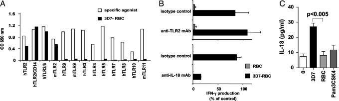
IFN-gamma secretion by natural killer (NK) cells is pivotal to several tumor and viral immune responses, during which NK and dendritic cells cooperation is required. We show here that macrophages are mandatory for NK cell IFN-gamma secretion in response to erythrocytes infected with Plasmodium falciparum (Pf), a causative agent of human malaria. In addition, direct sensing of Pf infection by NK cells induces their production of the proinflammatory chemokine CXCL8, without triggering their granule-mediated cytolytic programs. Despite their reported role in Pf recognition, Toll-like receptor (TLR) 2, TLR9, and TLR11 are individually dispensable for NK cell activation induced by Pf-infected erythrocytes. However, IL-18R expression on NK cells, IL-18 production by macrophages, and MyD88 on both cell types are essential components of this previously undescribed pathway of NK cell activation in response to a parasite infection.


Figures

Fig. 1.
Fig. 1.
Pf-RBC-induced NK cell activation within PBMC. Freshly isolated human PBMC were cultured alone (0) or in the presence of the NK cell target K562, Pf-RBC, or uninfected RBC (RBC). NK cell activation was analyzed by flow cytometry after gating on CD3-CD56+ NK cells (A-C) or on CD56+ lymphocytes (D). Statistical analyses were done by using a one-tailed Wilcoxon signed rank test. (A) After 20-24 h of coculture, the percentage of CD25+ cells within NK cells (Left) and the mean fluorescence intensity (MFI) of CD69 staining on NK cells (Right) are indicated for 30 healthy donors. Each dot represents the result obtained from one donor. (B) IFN-γ production by NK cells in PBMC was assessed by flow cytometry after coculture with 3D7-RBC or RBC during 20-24 h (Left) or 4 h in the presence of suboptimal doses of IL-12 (Right). Each dot represents the results from one donor. (C) IFN-γ production by NK cells was determined after 4 h of PBMC exposure to RBC infected with various Pf strains in the presence of IL-12. For each experiment, NK cell activation with the 3D7 strain was used as a control and represents 100% of NK cell IFN-γ production. Depending on the strains, 2-14 experiments were performed. Means ± SEM are represented. (D) Lamp1 and Lamp2 mobilization by NK cells (CD3-) or CD56+ T cells (CD3+) in PBMC was assessed by flow cytometry after 4 h of culture in the presence of monensin. One representative experiment of five is shown.
Fig. 2.
Fig. 2.
Full NK cell activation induced by Pf-RBC requires monocytes/macrophages. (A) Purified or FACS-sorted NK cells were exposed to medium alone (0), 3D7-RBC, or RBC for 24 h. (Upper) CD69 expression by NK cell assessed by flow cytometry (mean ± SEM of 10 experiments). (Lower) CXCL8 production in the supernatant of culture (mean ± SEM of 3 experiments). (B) After 4 h of exposure to RBC infected with 3D7 strain, in the presence of suboptimal doses of IL-12, the frequency of IFN-γ-producing NK cells was assessed by flow cytometry. In each experimental condition, the IFN-γ production was calculated as the proportion of IFN-γ-producing NK cells subtracted from background, as compared with IFN-γ-producing NK cells within PBMC in the presence of Pf-RBC (this “100% value” corresponds to 8.4% in this representative experiment). Purified NK cells were cultured alone or together with purified autologous DC, plasmacytoid DC (Upper) or FACS-sorted monocytes (Lower). CD14+ monocytes were also depleted from PBMC by FACS sorting before stimulation (Lower, NK in PBMC w/o monocytes). One representative experiment of three is shown.
Fig. 3.
Fig. 3.
Mouse NK cells are activated by Pf-RBC via an ITAM-independent and MyD88-dependent pathway. (A and B) Freshly isolated mouse splenocytes (from WT or genetically deficient mice for the indicated molecules) were cocultured with Pf-RBC (3D7-RBC) or uninfected RBC (RBC) for 20-24 h. NK cell activation was analyzed by flow cytometry after gating on CD3-NK1.1+ NK cells. Each dot represents the result from one individual mouse. Statistical analyses were done by using a one-tailed Wilcoxon signed rank test. (A Left and B) NK cell IFN-γ production was determined by intracytoplasmic staining after activation in the presence of suboptimal doses of IL-12. (A Right) CD69 expression on WT NK cell surface was assessed after indicated stimulation in the absence of exogenous cytokines (B) shows the percentage of IFN-γ-producing NK cells in the presence of 3D7-RBC (the background signals detected in the presence of uninfected RBC were subtracted).
Fig. 4.
Fig. 4.
MyD88 is required for NK cell activation induced by Pf-RBC. (A) Splenocytes from Ly5.1+ WT mice were mixed with splenocytes from either Ly5.1- MyD88-/- mice (Top), Ly5.1- IL-18R-/- mice (Middle), or Ly5.1- WT mice (Bottom) at a 1/1 ratio. Mixed splenocytes were then stimulated for 20-24 h in the presence of IL-12 with 3D7-RBC, RBC, or PMA/ionomycin. Two parameter flow cytometry contour plots, gated on NK cells, are shown. The frequencies of IFN-γ+ NK cells expressing Ly5.1+ or Ly5.1- are indicated in the upper and lower quadrant of each plot, respectively. Similar results were obtained in three experiments. (B) The Ly5.1+ mouse NK cell line Ky.2 was cultured in the absence (0) or presence of freshly isolated splenocytes from Ly5.1- WT or deficient mice and stimulated with 3D7-RBC for 20-24 h in the presence of IL-12. The percentages of Ly5.1+ Ky.2 cells producing IFN-γ are indicated. Results are expressed as means ± SEM of three experiments. Statistical analyses were performed by using a one-tailed Wilcoxon signed rank test. When cocultured with uninfected RBC, the percentage of IFN-γ+ Ky.2 cells was 1.9 ± 1.5 in the absence of splenocytes (n = 4), 4.4 ± 1.2 with WT splenocytes (n = 5), 2.9 ± 1.9 with MyD88 knockout (KO) splenocytes (n = 5), and 6.8 ± 2.0 with IL-18R knockout splenocytes (n = 3).
Fig. 5.
Fig. 5.
Human TLR2 is not mandatory for NK cell activation induced by Pf-RBC, but IL-18 is required. (A) HEK293 reporter cells transfected with indicated human (h) or mouse (m) TLR were stimulated with a specific agonist for each TLR (open bars) or 3D7-RBC (filled bars) for 18-24 h. Results are represented as optical density (OD) after subtraction of the background signal obtained with RBC or medium alone. (B) The production of IFN-γ by NK cells within human PBMC in response to 3D7-RBC versus RBC was assessed in the presence of anti-TLR2 blocking mAb, compared with the isotype control (Upper, n = 5), and in the presence of anti-IL-18 blocking mAb, compared with the isotype control (Lower, n = 2). Results (means ± SD) are presented as a percentage of control calculated as in Fig. 2. (C) IL-18 production was assayed in the supernatant of purified human macrophages after 24 h of culture with Pam3CSK4, 3D7-RBC, or RBC, in the presence of medium alone.

References

    1. Dokun, A. O., Chu, D. T., Yang, L., Bendelac, A. S. & Yokoyama, W. M. (2001) J. Immunol. 167, 5286-5293. - PubMed
    1. Walzer, T., Dalod, M., Robbins, S. H., Zitvogel, L. & Vivier, E. (2005) Blood 106, 2252-2258. - PubMed
    1. Moretta, L. & Moretta, A. (2004) EMBO J. 23, 255-259. - PMC - PubMed
    1. Raulet, D. H. (2004) Nat. Immunol. 5, 996-1002. - PubMed
    1. Degli-Esposti, M. A. & Smyth, M. J. (2005) Nat. Rev. Immunol. 5, 112-124. - PubMed

Publication types

MeSH terms