Comparative Study
. 2005 Oct 11;102(41):14677-82.
doi: 10.1073/pnas.0507250102. Epub 2005 Oct 3.

Long-term renewal of hair follicles from clonogenic multipotent stem cells

Affiliations
Comparative Study

Long-term renewal of hair follicles from clonogenic multipotent stem cells

Stéphanie Claudinot et al. Proc Natl Acad Sci U S A. .

Abstract

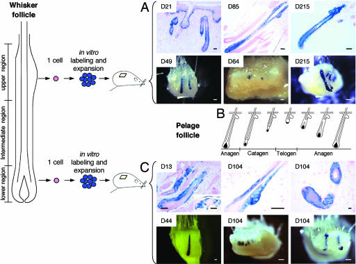
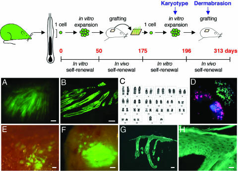
Adult stem cells are essential for tissue renewal, regeneration, and repair, and their expansion in culture is of paramount importance for regenerative medicine. Using the whisker follicle of the rat as a model system, we demonstrate that (i) clonogenicity is an intrinsic property of the adult stem cells of the hair follicle; (ii) after cultivation for >140 doublings, these stem cells, transplanted to the dermo-epidermal junction of newborn mouse skin, form part or all of the developing follicles; (iii) the stem cells incorporated into follicles are multipotent, because they generate all of the lineages of the hair follicle and sebaceous gland; (iv) thousands of hair follicles can be generated from the progeny of a single cultivated stem cell; (v) cultured stem cells express the self-renewal genes Bmi1 and Zfp145;(vi) several stem cells participate in the formation of a single hair bulb; and (vii) there are many more stem cells in whisker follicles than could be anticipated from label-retaining experiments.


Figures

Fig. 1.
Fig. 1.
Clonogenic keratinocytes are long-term multipotent stem cells. Single keratinocytes were isolated from the upper or lower regions of whisker follicles of adult rats. Clones were labeled by using a defective retrovirus bearing a β-gal gene and subcloned, and labeled cells (passages 11-12) were injected into newborn mouse skin (Fig. 6). Transplanted clonogenic keratinocytes, whether from the upper or lower regions, contributed to all epithelial lineages of hairy skin, including sebaceous glands, and participated in the hair cycle of pelage follicles for months. Note the variable degree of chimerism and that the epidermis is not labeled in long-term grafts (Lower in A and C). Insets show duration of transplantation in days in A and C. D21-D85 follicles were from subclone RA15 of clone YR25P3-cl4; D215 follicles were from subclone RA9 of clone YR25P3-cl4. D49 (anagen) and D64 (early anagen) follicles were from subclone RA15 of clone YR25P3-cl4. D215 follicles (anagen and telogen) were from subclone RA9 of clone YR25P3-cl4. In Lower, all follicles were from clone YR207P1-cl16. (B) Schematic representation of the hair cycle phases of a pelage follicle. (Scale bars: biopsies stained for β-gal in toto, 100 μm; microscopic sections, 50 μm.)
Fig. 2.
Fig. 2.
Long-term persistence of hair follicles generated from a single multipotent stem cell. A single cell (clone YR219P3-cl7) was directly isolated from the upper region of a whisker follicle of an EGFP rat. After cultivation for 50 days, it was transplanted. One hundred twenty-five days later, a biopsy was obtained from the graft, and clonogenic EGFP+ cells were isolated, subcloned, and cultivated for 3 weeks before they were again transplanted. (A) UV illumination of a biopsy 117 days after the second transplantation; numerous EGFP+ follicles were present. The epidermis was not labeled. (B) Frozen section of A. (C) Cells of subclone 14 of clone YR219P3-cl7 were diploid (2n = 42). (D) FISH analysis shows that no fusion occurred between rat (green probe) and mouse (red probe) cells during the transplantation; cells labeled in red are irradiated 3T3 feeder cells. Nuclei are blue (DAPI). (E) Graft of subclone 14 of clone YR219P3-cl7 at 54 days after the second transplantation. Labeled follicles appeared as green spots under UV illumination. The epidermis was not labeled. (F) The same graft 4 days after dermabrasion with sandpaper. The surface of the graft was bright green (compare with A). (G) Microscopic appearance of F. An EGFP+ epidermis formed, indicating that EGFP+ cells moved out of the hair follicles. (H) Detail is shown. (Scale bars: A and E-G, 100 μm; B, 50 μm; H, 20 μm.)
Fig. 3.
Fig. 3.
Homing to the niche. (A) Location of the niche (orange) in a pelage follicle below the sebaceous gland. (B) Transplanted stem cells (subclone RA15 of clone YR25P3-cl4) correctly homed to the bulge region (arrow) of a mouse pelage hair follicle 64 days after transplantation. The follicle shown was in early anagen. Note the sharp boundary between the follicle and the unlabeled epidermis. Note also that the sebaceous glands and the lower part of the infundibulum contain labeled cells. (C) Labeled stem cells intermingled with unlabeled resident stem cells (arrow). (D) The hair germ was mostly constituted of labeled cells; the dermal papilla cells located below were unlabeled. (Scale bars: B, 50 μm; C and D, 20 μm.)
Fig. 4.
Fig. 4.
Mosaicism indicates that the hair bulb is polyclonal. (Left) Schematic drawing of cross sections of an anagen follicle modified from ref. . (A-D) Labeled follicles were often mosaic, indicating that several stem cells participated in their formation or renewal (see Figs. 1 and 3). Mosaicism was often observed within a single follicular sheath, indicating that the committed progenitors that participated in the formation of that particular sheath were generated by different stem cells. (Scale bars: 20 μm.)
Fig. 5.
Fig. 5.
Cultivated stem cells express Bmi1 and Zfp145. Clones were obtained from the upper and lower regions of whisker follicles of EGFP rats (clones YR219P5-cl7 and YR221P1-cl10.3). EGFP+ cells were sorted from 3-day-old cultures at passages 7 and 11 to eliminate the irradiated 3T3 cells. mRNAs were then extracted, and RT-PCRs were performed using specific primers (see Table 1). Mouse ES cells were used as a positive control for the expression of Oct-4, Bmi1, and Zfp145.

Similar articles

Cited by

References

    1. Watt, F. M. & Hogan, B. L. (2000) Science 287, 1427-1430. - PubMed
    1. Gambardella, L. & Barrandon, Y. (2003) Curr. Opin. Cell Biol. 15, 771-777. - PubMed
    1. Fuchs, E., Tumbar, T. & Guasch, G. (2004) Cell 116, 769-778. - PubMed
    1. Schofield, R. (1978) Blood Cells 4, 7-25. - PubMed
    1. Nilsson, S. K. & Simmons, P. J. (2004) Curr. Opin. Hematol. 11, 102-106. - PubMed

Publication types

MeSH terms

LinkOut - more resources