

Corroboration of in vivo cartilage pressures with implications for synovial joint tribology and osteoarthritis causation
- PMID: 16203974
- PMCID: PMC1253592
- DOI: 10.1073/pnas.0507117102
Corroboration of in vivo cartilage pressures with implications for synovial joint tribology and osteoarthritis causation
Abstract
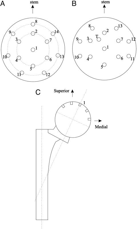
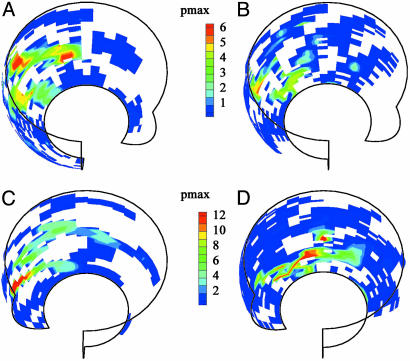
Pressures on normal human acetabular cartilage have been collected from two implanted instrumented femoral head hemiprostheses. Despite significant differences in subjects' gender, morphology, mobility, and coordination, in vivo pressure measurements from both subjects covered similar ranges, with maximums of 5-6 MPa in gait, and as high as 18 MPa in other movements. Normalized for subject weight and height (nMPa), for free-speed walking the maximum pressure values were 25.2 for the female subject and 24.5 for the male subject. The overall maximum nMPa values were 76.2 for the female subject during rising from a chair at 11 months postoperative and 82.3 for the male subject while descending steps at 9 months postoperative. These unique in vivo data are consistent with corresponding cadaver experiments and model analyses. The collective results, in vitro data, model studies, and now corroborating in vivo data support the self-pressurizing "weeping" theory of synovial joint lubrication and provide unique information to evaluate the influence of in vivo pressure regimes on osteoarthritis causation and the efficacy of augmentations to, and substitutions for, natural cartilage.
Figures


References
-
- Kuettner, K. E., Schleyerbach, R., Peyron, J. G. & Hascall, V. C., eds. (1991) Articular Cartilage and Osteoarthritis (Raven, New York).
-
- Shakoor, N. & Loeser, R. F. (2004) Sci. Aging Knowl. Environ. 35, dn2. - PubMed
-
- National Institute of Arthritis and Musculoskeletal and Skin Diseases, National Institutes of Health (2005) Cartilage and Connective Tissue Research, www.niams.nih.gov/an/stratplan/cartilage.htm.
-
- Sokoloff, L. (1969) The Biology of Degenerative Joint Disease (Univ. of Chicago Press, Chicago).
-
- Radin, E. L., Ehrlich, M. G. & Chernack, R. (1978) Clin. Orth. 131, 288-293. - PubMed
Publication types
MeSH terms
Grants and funding
LinkOut - more resources
Full Text Sources
Other Literature Sources
Medical
