

doi: 10.1101/gad.1339805.
Cdc7-Drf1 is a developmentally regulated protein kinase required for the initiation of vertebrate DNA replication
Affiliations
- PMID: 16204181
- PMCID: PMC1240038
- DOI: 10.1101/gad.1339805
Item in Clipboard
Cdc7-Drf1 is a developmentally regulated protein kinase required for the initiation of vertebrate DNA replication
Genes Dev.
.
Abstract
Cdc7, a protein kinase required for the initiation of eukaryotic DNA replication, is activated by a regulatory subunit, Dbf4. A second activator of Cdc7 called Drf1 exists in vertebrates, but its function is unknown. Here, we report that in Xenopus egg extracts, Cdc7-Drf1 is far more abundant than Cdc7-Dbf4, and removal of Drf1 but not Dbf4 severely inhibits phosphorylation of Mcm4 and DNA replication. After gastrulation, when the cell cycle acquires somatic characteristics, Drf1 levels decline sharply and Cdc7-Dbf4 becomes the more abundant kinase. These results identify Drf1 as a developmentally regulated, essential activator of Cdc7 in Xenopus.
Figures
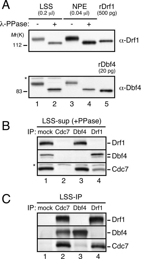
Cdc7-Drf1 is far more abundant than Cdc7-Dbf4 in Xenopus egg extracts. (A) Characterization of anti-Drf1 and Dbf4 antibodies: 0.2 μL LSS (lane 1), 0.2 μL λ-protein phosphatase-treated LSS (lane 2), 0.04 μL NPE (lane 3), and 0.04 μL λ-protein phosphatase-treated NPE (lane 4) were separated by SDS-PAGE and probed with anti-Drf1 (top panel) or Dbf4 (bottom panel) antibodies along-side 500 pg of recombinant Drf1 (6xHis and StrepII-tagged) (lane 5, top panel) or 20 pg recombinant Dbf4 (6xHis-tagged) (lane 5, bottom panel). Asterisks indicate cross-reacting bands. (B,C) Cdc7 complexes were immunoprecipitated from LSS by control (lane 1), anti-Cdc7 (lane 2), anti-Dbf4 (lane 3), or anti-Drf1 (lane 4) antibodies, and the supernatant (B) and immunoprecipitate (C) fractions were probed with antibodies against Drf1 (top panels), Dbf4 (middle panels), and Cdc7 (bottom panels). Cross-reacting bands (*) serve as loading controls. In B and later figures involving Dbf4 blotting of LSS, extracts were treated with phosphatase due to a cross-reacting band migrating with phosphorylated Dbf4.
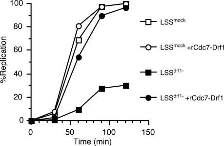
Drf1 is required for DNA replication. Sperm chromatin was incubated in mock-depleted LSS supplemented with buffer (open squares) or 40 nM rCdc7-Drf1 (open circles), or Drf1-depleted LSS supplemented with buffer (filled squares) or 40 nM rCdc7-Drf1 (filled circles).
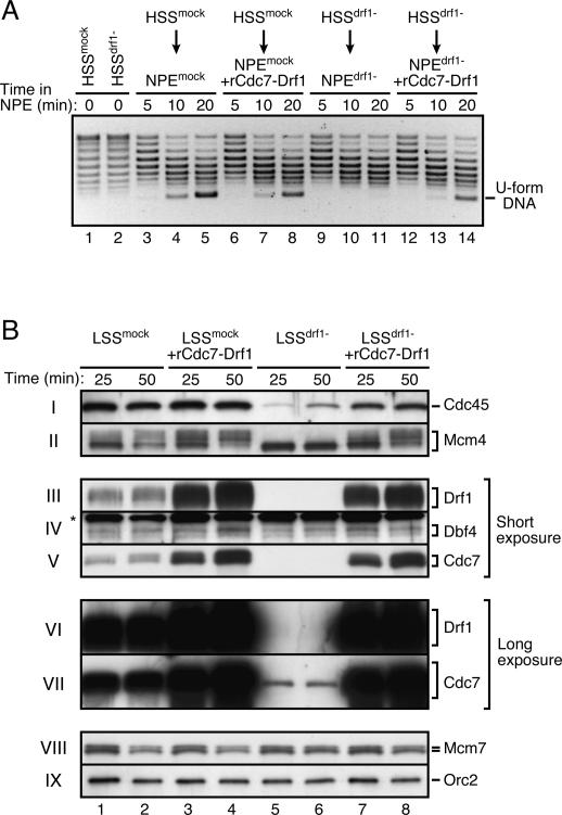
Cdc7-Drf1 is required for initiation of DNA replication. (A) Drf1 is required for origin unwinding. pBluescriptII was incubated in mock-depleted (lane 1) or Drf1-depleted (lane 2) HSS at 40 ng/μL concentration for 30 min and isolated, or further incubated in mock-depleted (lanes 3-8) or Drf1-depleted (lanes 9-14) NPE containing 50 μg/mL aphidicolin and buffer (lanes 3-5,9-11) or 500 nM rCdc7-Drf1 (lanes 6-8,12-14). At the indicated times, the plasmid topology was analyzed by agarose gel electrophoresis in the presence of 1.8 μM chloroquine. (B) Requirement of Drf1 for Mcm4 phosphorylation and Cdc45 loading. Sperm chromatin was incubated in mock-depleted (lanes 1-4) or Drf1-depleted (lanes 5-8) LSS containing buffer (lanes 1,2,5,6) or 50 nM rCdc7-Drf1 (lanes 3,4,7,8) and recovered through a sucrose cushion at the indicated time points. Bound proteins were eluted and analyzed by Western blotting using the indicated antibodies. (*) Cross-reacting band.
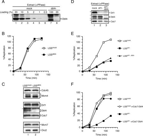
Dbf4 is not required for DNA replication in Xenopus egg extracts. (A) The indicated amounts of mock-depleted (lanes 1-6) or Dbf4-depleted (lanes 7,8) LSS treated with λ-protein phosphatase were separated on SDS-PAGE and probed with anti-Dbf4 antibody; 100% = 1 μL. Cross-reacting bands (*) serve as loading controls. (B) Replication efficiency in the mock-depleted (open squares) or Dbf4-depleted (filled squares) LSS shown in A. (C) Sperm chromatin was incubated in mock-depleted (lane 1) or Dbf4-depleted (lane 2) LSS in the presence of 50 μg/mL aphidicolin and isolated after 45 min. Western blotting for the indicated proteins is presented. (*) Cross-reacting band. In D, 0.2 μL each of mock-depleted (lane 1), Drf1-depleted (lane 2), or Drf1/Dbf4-depleted (lane 3) LSS was treated with λ-protein phosphatase, separated by SDS-PAGE, and probed for the indicated proteins. (*) Cross-reacting band. (E) Replication efficiency in mock-depleted (open squares), Drf1-depleted (filled squares), and Drf1- and Dbf4-depleted (filled triangles) LSS shown in D.(F) Replication efficiency in LSS that was mock-depleted (open squares), Drf1-depleted (filled squares), mock-depleted and supplemented with 1 μM recombinant Cdc7-Dbf4 (open circles), and Drf1-depleted and supplemented with 1 μM recombinant Cdc7-Dbf4 (filled circles).
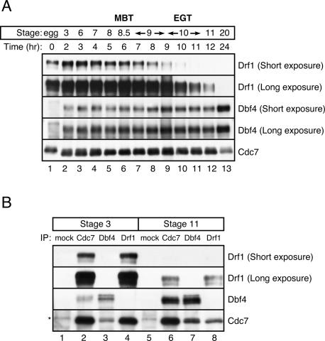
Developmental regulation of Drf1 and Dbf4. (A) Unfertilized Xenopus eggs (lane 1) or in vitro fertilized Xenopus embryos (lanes 2-13) were isolated at the indicated time points and staged according to Nieuwkoop and Faber (1967). Cdc7 complexes were immunoprecipitated from embryo lysates with anti-Cdc7 antibody and probed for Drf1 (top and second panels), Dbf4 (third and fouth panels), and Cdc7 (bottom panel). (B) Stage 3 in vitro fertilized Xenopus embryos (lanes 1-4) or stage 11 embryos (lanes 5-8) were collected and used for immunoprecipitation with control (lanes 1,5), anti-Cdc7 (lanes 2,6), anti-Dbf4 (lanes 3,7), and anti-Drf1 (lanes 4,8) antibodies. Western blotting for the indicated proteins is presented. An asterisk shows the IgG heavy chain, which reacts with the secondary antibody.
Similar articles
-
Xenopus CDC7/DRF1 complex is required for the initiation of DNA replication.J Biol Chem. 2006 Apr 28;281(17):11569-76. doi: 10.1074/jbc.M510278200. Epub 2006 Feb 28. J Biol Chem. 2006. PMID: 16507577
-
Drf1-dependent kinase interacts with Claspin through a conserved protein motif.J Biol Chem. 2010 Apr 23;285(17):12638-46. doi: 10.1074/jbc.M109.077370. Epub 2010 Feb 27. J Biol Chem. 2010. PMID: 20190277 Free PMC article.
-
DBF4, not DRF1, is the crucial regulator of CDC7 kinase at replication forks.J Cell Biol. 2024 Aug 5;223(8):e202402144. doi: 10.1083/jcb.202402144. Epub 2024 Jun 12. J Cell Biol. 2024. PMID: 38865090 Free PMC article.
-
Cdc7 kinase complex: a key regulator in the initiation of DNA replication.J Cell Physiol. 2002 Mar;190(3):287-96. doi: 10.1002/jcp.10070. J Cell Physiol. 2002. PMID: 11857444 Review.
-
Regulation and roles of Cdc7 kinase under replication stress.Cell Cycle. 2014;13(12):1859-66. doi: 10.4161/cc.29251. Epub 2014 May 19. Cell Cycle. 2014. PMID: 24841992 Free PMC article. Review.
Cited by
-
Evolutionary diversification of MCM3 genes in Xenopus laevis and Danio rerio.Cell Cycle. 2014;13(20):3271-81. doi: 10.4161/15384101.2014.954445. Cell Cycle. 2014. PMID: 25485507 Free PMC article.
-
Integrating S-phase checkpoint signaling with trans-lesion synthesis of bulky DNA adducts.Cell Biochem Biophys. 2007;47(3):392-408. doi: 10.1007/s12013-007-0032-7. Cell Biochem Biophys. 2007. PMID: 17652783 Free PMC article. Review.
-
Biphasic chromatin binding of histone chaperone FACT during eukaryotic chromatin DNA replication.Biochim Biophys Acta. 2011 Jun;1813(6):1129-36. doi: 10.1016/j.bbamcr.2011.01.002. Epub 2011 Jan 11. Biochim Biophys Acta. 2011. PMID: 21232560 Free PMC article.
-
Chk1 Inhibition of the Replication Factor Drf1 Guarantees Cell-Cycle Elongation at the Xenopus laevis Mid-blastula Transition.Dev Cell. 2017 Jul 10;42(1):82-96.e3. doi: 10.1016/j.devcel.2017.06.010. Dev Cell. 2017. PMID: 28697335 Free PMC article.
-
The MCM2-7 Complex: Roles beyond DNA Unwinding.Biology (Basel). 2024 Apr 13;13(4):258. doi: 10.3390/biology13040258. Biology (Basel). 2024. PMID: 38666870 Free PMC article. Review.
References
-
- Bell S.P. and Dutta, A. 2002. DNA replication in eukaryotic cells. Annu. Rev. Biochem. 71: 333-374. - PubMed
-
- Blow J.J. and Laskey, R.A. 1986. Initiation of DNA replication in nuclei and purified DNA by a cell-free extract of Xenopus eggs. Cell 47: 577-587. - PubMed
-
- Brown G.W. and Kelly, T.J. 1998. Purification of Hsk1, a minichromosome maintenance protein kinase from fission yeast. J. Biol. Chem. 273: 22083-22090. - PubMed
-
- Costanzo V., Shechter, D., Lupardus, P.J., Cimprich, K.A., Gottesman, M., and Gautier, J. 2003. An ATR- and Cdc7-dependent DNA damage checkpoint that inhibits initiation of DNA replication. Mol. Cell 11: 203-213. - PubMed
Publication types
MeSH terms
Substances
Grants and funding
LinkOut - more resources
Full Text Sources