. 2006 Jan;172(1):445-56.
doi: 10.1534/genetics.105.048934. Epub 2005 Oct 3.

A novel murine gene, Sickle tail, linked to the Danforth's short tail locus, is required for normal development of the intervertebral disc

Affiliations

A novel murine gene, Sickle tail, linked to the Danforth's short tail locus, is required for normal development of the intervertebral disc

Kei Semba et al. Genetics. 2006 Jan.

Abstract

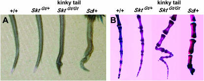
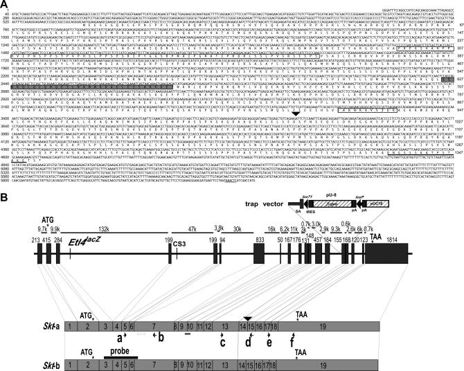
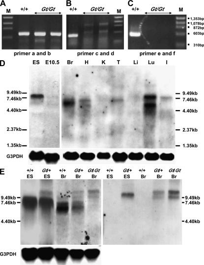
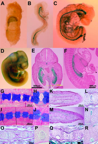
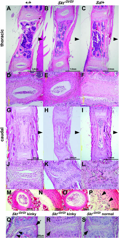
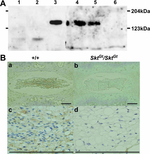
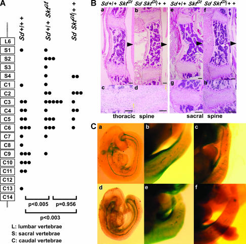
We established the mutant mouse line, B6;CB-SktGtAyu8021IMEG (SktGt), through gene-trap mutagenesis in embryonic stem cells. The novel gene identified, called Sickle tail (Skt), is composed of 19 exons and encodes a protein of 1352 amino acids. Expression of a reporter gene was detected in the notochord during embryogenesis and in the nucleus pulposus of mice. Compression of some of the nuclei pulposi in the intervertebral discs (IVDs) appeared at embryonic day (E) 17.5, resulting in a kinky-tail phenotype showing defects in the nucleus pulposus and annulus fibrosus of IVDs in SktGt/Gt mice. These phenotypes were different from those in Danforth's short tail (Sd) mice in which the nucleus pulposus was totally absent and replaced by peripheral fibers similar to those seen in the annulus fibrosus in all IVDs. The Skt gene maps to the proximal part of mouse chromosome 2, near the Sd locus. The genetic distance between them was 0.95 cM. The number of vertebrae in both [Sd +/+ SktGt] and [Sd SktGt/+ +] compound heterozygotes was less than that of Sd heterozygotes. Furthermore, the enhancer trap locus Etl4lacZ, which was previously reported to be an allele of Sd, was located in the third intron of the Skt gene.


Figures

Figure 1.
Figure 1.
(A) Tail phenotype of 8-week-old mice. SktGt/Gt mice had kinked tails compared to wild-type, SktGt/+, and heterozygous Sd mice. (B) Alizarin red whole-mount preparations of the tails of 8-week-old mice. SktGt/Gt mice confirmed this kinky-tail phenotype. Sd mice showed decreased numbers of vertebrae with truncation at the caudal vertebrae.
Figure 2.
Figure 2.
Identification of the trapped gene Skt. (A) The nucleotide sequence of the Skt cDNA and predicted amino acid sequence. The open box indicates the pro-rich region at the N terminus and the shaded box indicates the coiled-coil region in the middle. The striped box indicates the sequence deleted in Skt-b by alternative splicing. The 15-amino-acid peptide used for the production of anti-Skt antibodies is shown by underlining. The polyadenylation signal is underlined at the 3′-end of the nucleotide sequence. The nucleotide sequence is numbered on the left side and the amino acid sequence is numbered on the right side. (B) Genomic structure of the Skt gene. (Top) Exon-intron structure of the Sickle tail gene. The trap vector, pU-8, was inserted into the 14th intron. Sizes of exons and introns are given. (Bottom) Two transcripts produced from the Skt allele. There are at least two types of Skt transcripts: one contains all the exons (termed Skt-a) and the other lacks 33 bp of the 13th exon (termed Skt-b). Arrows (a–f) indicate the location of the primers used for RT–PCR analyses in Figure 3, A–C, to detect the expression of each part of the Skt transcripts. The solid bar represents a probe used for Northern blotting. The open and shaded boxes indicate the pro-rich region and the coiled-coil region, respectively. The start and stop codons of the Skt gene are shown by asterisks. A sequence with high homology to the CS3 in node/notochord enhancers is located in the fourth intron of the Skt gene 106 kb downstream of the insertion site of Etl4lacZ.
Figure 3.
Figure 3.
Analyses of Skt transcripts. (A–C) RT–PCR analyses using E10.5 embryos to detect Skt transcripts in wild-type (+/+) and SktGt/Gt embryos. The transcripts containing nucleotide sequences upstream of the insertion site of the trap vector were detected in both wild-type and SktGt/Gt embryos (A). The transcripts containing nucleotide sequences downstream of the Skt sequence were not detected in SktGt/Gt embryos (B and C). M, molecular marker. (D) Northern blot analyses to detect Skt mRNA in the wild-type ES cells, E10.5 embryo, and 8-week-old mice, using the Skt-specific probe in the 5′-region (see Figure 2B). Total RNA (10 μg) from TT2 ES cells, wild-type E10.5 embryos, mRNA (5 μg) from wild-type organs, and a Skt RNA probe were used for Northern blotting. (E) Northern blot analyses to detect Skt and β-geo fusion transcripts. Total RNA (20 μg) from TT2 and heterozygous (Gt/+) ES cells, wild-type (+/+), heterozygous (Gt/+), and homozygous (Gt/Gt) adult brains was used for Northern blotting. The Skt RNA probe or lacZ RNA probe was used in the left and right panels, respectively. Br, brain; H, heart; K, kidney; T, testis; Li, liver; Lu, lung; I, intestine.
Figure 4.
Figure 4.
β-gal expression and histological analyses in SktGtmice. At E7.5 (A), the chorion was stained with X-gal, but the midline region of the embryo was not stained. At E8.5 (B) and E9.0 (C) intense staining was detected in the notochord. At E11.5 (D–F), the notochord and the mesonephros expressed β-geo strongly in whole-mount X-gal staining (D), the frontal section (E), and the sagittal section (F). Sagittal sections of the tail bud of SktGt/+ (G and H) and SktGt/Gt (I and J) embryos at E17.5. At E17.5, some IVDs were compressed in the tail bud of SktGt/Gt (J). Sagittal sections of the tail tips of newborn SktGt/+ (K and L) and SktGt/Gt (M and N) mice. In the SktGt/Gt neonate, the vertebral body alignment was undulated (M and N). Sagittal sections of the tail tips of 2-week-old SktGt/+ (O and P) and SktGt/Gt (Q and R) mice. In the 2-week-old SktGt/Gt mice, the X-gal-positive nuclei pulposi were dislocated to the periphery (Q and R). (H, J, L, N, P, and R) Higher magnification of the area indicated by the boxes in G, I, K, M, O, and Q, respectively. Sections (G–J) were stained with alcian blue and sections of X-gal staining (E, F, and K–R) were counterstained with Nuclear Fast red. Bars, 200 μm.
Figure 5.
Figure 5.
Histological analyses of IVDs of SktGt/Gt and Sd mutant adult mice. Sagittal sections of the thoracic and caudal IVD from 8-week-old adult wild-type (A, D, G, and J), SktGt/Gt (B, E, H, and K), and heterozygous Sd mice (C, F, I, and L). (D, E, F, J, K, and L) Higher magnification of the area indicated by the boxes in A, B, C, G, H, and I, respectively. Arrowheads indicate the dorsal side. Axial sections of the upper caudal IVD in wild-type (M and N) and SktGt/Gt (O and P) 8-week-old mice. (N and P) Higher magnification of the area indicated by the boxes in M and O, respectively. The arrowheads in P indicate an irregular boundary with close contact between the nucleus pulposus and annulus fibrosus. (Q–S) The 20–25th caudal IVDs of SktGt/Gt 8-week-old mice. Impaired development of the annulus fibrosus in SktGt/Gt mice was demonstrated by the thin fibrous layers of annulus fibrosus (Q and R) and the failure of fibrous adhesion to the vertebral bodies (arrowhead in Q). Similar IVD abnormalities such as dislocation of the nucleus pulposus (arrow in Q–S) and impaired growth of the annulus fibrosus were observed in the nonkinked regions (S) and in the kinked regions (Q and S). Haematoxylin and eosin (HE) staining was used. Bars, 200 μm.
Figure 6.
Figure 6.
Detection of the Skt protein. (A) Western blot analysis to detect Skt protein using extracts from untreated BMT10 cells (lane 1), BMT10 cells transfected with vector (lane 2), BMT10 cells transfected with the Skt expression vector (lane 3), and extracts of the nucleus pulposus of caudal IVDs from 8-week-old wild-type mice (lane 4), SktGt/+ mice (lane 5), and SktGt/Gt mice (lane 6). An ∼150-kDa protein corresponding to the predicted molecular weight of 147 kDa was detected in lanes 3, 4, and 5, but not in lane 6. The amount of Skt protein was reduced in the SktGt/+ mutant (lane 5) and was below the detectable level in SktGt/Gt (lane 6). (B) Immunohistochemistry of frontal sections of the nucleus pulposus in upper caudal IVDs from adult 8-week-old mice using purified anti-Skt antibodies. Skt protein was detected in the cytoplasm of nucleus pulposus cells in wild-type (a and c), but not SktGt/Gt, mice (b and d). (c and d) Higher magnification of the area indicated by the boxes in a and b, respectively. Bars, 200 μm.
Figure 7.
Figure 7.
(A) Schematic of axial levels and severity of vertebral malformations in compound mutant 8-week-old mice. A single solid circle indicates the level of the terminal vertebral body for a single mouse. In both trans and cis compound mutant mice, the degree of vertebral malformation is more severe than that in heterozygous Sd mice (trans, P < 0.005; cis, P < 0.003, Mann–Whitney U-test). There was no significant difference between trans [Sd +/+ SktGt] (n = 23) and cis [Sd SktGt/+ +] (n = 10) mice (P = 0.956, Mann–Whitney U-test). (B) Histological analyses of vertebral columns in the Sd +/+ SktGt and Sd SktGt/+ + mutant mice. HE staining of midsagittal sections in thoracic spines of Sd +/+ SktGt (a and c) and Sd SktGt /+ + (b and d) mice and in sacral spines of Sd +/+ SktGt (e and g) and Sd SktGt/+ + (f and h) mice. Arrowheads indicate dorsal sides. (c, d, g, and h) Higher magnification of the areas indicated by the boxes in a, b, e, and f, respectively. (C) Whole-mount X-gal staining in the Sd +/+ SktGt and Sd SktGt/+ + mutant embryos. The β-gal expression in the tail notochord of trans-heterozygous embryos [Sd +/+ SktGt] at E9.5 (a) and E13.5 (b and c) and of cis-heterozygous embryos [Sd SktGt/+ +] at E9.5 (d) and E13.5 (e and f).

Similar articles

Cited by

References

    1. Alfred, J. B., K. Rance, B. A. Taylor, S. J. Phillips, C. M. Abbott et al., 1997. Mapping in the region of Danforth's short tail and the localization of tail length modifiers. Genome Res. 7: 108–117. - PubMed
    1. Allen, N. D., D. G. Cran, S. C. Barton, S. Hettel, W. Reik et al., 1988. Transgenes as probes for active chromosomal domains in mouse development. Nature 333: 852–855. - PubMed
    1. Ang, S., and J. Rossant, 1994. HNF-3β is essential for node and notochord formation in mouse development. Cell 78: 561–574. - PubMed
    1. Araki, K., T. Imaizumi, T. Sekimoto, K. Yoshinobu, J. Yoshimuta et al., 1999. Exchangeable gene trap using the Cre/mutated lox system. Cell. Mol. Biol. 45: 737–750. - PubMed
    1. Aszodi, A., D. Chan, E. Hunziker, J. F. Bateman and R. Fassler, 1998. Collagen II is essential for the removal of the notochord and the formation of intervertebral discs. J. Cell Biol. 143: 1399–1412. - PMC - PubMed

Publication types

MeSH terms

Associated data