Comparative Study
. 2006 Jan;172(1):7-15.
doi: 10.1534/genetics.105.048553. Epub 2005 Oct 3.

Identification and analysis of Escherichia coli ribonuclease E dominant-negative mutants

Affiliations
Comparative Study

Identification and analysis of Escherichia coli ribonuclease E dominant-negative mutants

Karoline J Briegel et al. Genetics. 2006 Jan.

Abstract

The Escherichia coli (E. coli) ribonuclease E protein (RNase E) is implicated in the degradation and processing of a large fraction of RNAs in the cell. To understand RNase E function in greater detail, we developed an efficient selection method for identifying nonfunctional RNase E mutants. A subset of the mutants was found to display a dominant-negative phenotype, interfering with wild-type RNase E function. Unexpectedly, each of these mutants contained a large truncation within the carboxy terminus of RNase E. In contrast, no point mutants that conferred a dominant-negative phenotype were found. We show that a representative dominant-negative mutant can form mixed multimers with RNase E and propose a model to explain how these mutants can block wild-type RNase E function in vivo.


Figures
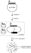
Figure 1.
Figure 1.
Strategy used to identify RNase E dominant-negative mutants. Plasmid pLAC-RNE contains an rne gene (shaded rectangle) transcribed from a Plac promoter. Plasmid DNA was mutagenized (materials and methods) and transformed into the reporter strain CJ1830. CJ1830 contains an rne gene encoding wild-type RNase E transcribed from a Plac promoter and a chromosomal rne-lacZ reporter fusion (shaded/solid rectangle) that is negatively regulated by RNase E. Because CJ1830 lacks Lac repressor, Plac expression of the plasmid encoded rne gene is fully induced. Therefore, the expression of the wild-type RNase E protein or of mutants that retain substantial activity causes cell death. Only transformants that contain RNase E mutants that display a significant defect are able to form viable colonies (denoted below as circles). Amongst these, a subset of the mutants that confer a dominant-negative phenotype show an enhanced lacZ phenotype (solid circles).
Figure 2.
Figure 2.
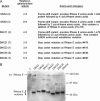
RNase E dominant-negative mutants isolated in this study. (A) Predicted amino acid changes and increase in rne-lacZ activity for the dominant-negative mutants. The mutant designations are indicated in the first column, with the number of times each mutant was isolated denoted in brackets. The second column indicates the fold increase in β-galactosidase activity when CJ1830 cells were transformed with the corresponding dominant-negative mutant. To derive these values, the β-galactosidase activity of each CJ1830 strain expressing the indicated dominant-negative mutant was divided by the β-galactosidase activity of CJ1830 containing the control plasmid, pACYC184 (typically, 80 Miller units; Miller 1972). Each measurement reflects the average of three independent determinations, with standard deviations typically <10%. The predicted amino acid changes for each RNase E variant is shown in the last column. (B) To confirm that the dominant-negative mutants encode truncated proteins, cell extracts derived from the strains used for the lacZ assays were used for Western blot analysis using anti-RNase E antibodies. The positions of the wild-type chromosomally encoded RNase E and the truncated plasmid-encoded dominant-negative mutants are indicated. The migration of the protein molecular weight markers is also shown. C, control strain containing pACYC184, a non-rne plasmid.
Figure 3.
Figure 3.
Expression of dominant-negative mutants in a wild-type rne strain background. (A) Plasmids containing the indicated mutants or a control plasmid (pACYC184) were transformed into CJ1825. The transformants were grown to midlog phase and assayed for β-galactosidase activity. The magnitude of the dominant-negative effect is the β-galactosidase activity of CJ1825 derivatives expressing the indicated dominant-negative mutant divided by the β-galactosidase activity of the same strain containing pACYC184. (B) Western blot analysis. Strain CJ1825 transformed with pACYC184 or pDNX5 was grown to midlog phase in LB plus Cm and used to prepare cell extracts. A total of 20 μg of cell-extract protein was fractionated by SDS-PAGE and analyzed by Western blot analysis using anti-RNase E antibodies. The migration of full-length RNase E is indicated by an arrow.
Figure 4.
Figure 4.
Effect of expressing the DNX5 RNase E variant on the half-life of rpsO and RNA I transcripts. CJ1825 transformed with pACYC184 or pDNX5 was grown to midlog phase, treated with rifampicin, and used to prepare total RNA from fractions harvested at different times. Equal amounts of each RNA sample were used for primer extension using rpsO and RNA I-specific probes and fractionated on a denaturing polyacrylamide gel alongside radiolabeled 100 base pair DNA markers (lane 1). The positions of the 85-nucleotide RNA I and the 266-nucleotide rpsO primer extension products are indicated. For each RNA sample used, the time (in minutes) at which the corresponding samples were harvested after rifampicin addition is indicated.
Figure 5.
Figure 5.
Complex formation between the DNX5 RNase E mutant and the His-tagged amino-terminal domain of RNase E. Plasmids pDNX5 and pNRNE5, which encode a His-tagged amino-terminal domain of RNase E were transformed individually or together into strain CJ1825. Transformed colonies were grown in LB medium containing the appropriate antibiotics and induced with IPTG prior to harvesting. (A) Equal amounts of cell lysates prepared from these strains were fractionated by SDS-PAGE, transferred to nitrocellulose membrane, and the RNase E products were detected by Western blot analysis using anti-RNase E antibodies. (B) Each of the cell lysates was incubated with Ni-NTA beads, after which the beads were washed extensively to remove unbound protein. Thereafter, His-tagged RNase E was eluted by adding buffer containing imidazole. Equal volumes of the eluted fractions were fractionated by PAGE and analyzed by Western blot analysis.
Figure 6.
Figure 6.
Model for the dominant-negative phenotype conferred by truncated RNase E variants. RNase E is proposed to bind RNA as a multimer, with two or more RNase E monomers in this complex contributing to RNA binding. For simplicity, RNase E is shown as a dimer, and only the catalytic domain and RNA-binding domains are depicted schematically. The recruitment of the RNA substrates to the enzyme-active site leads to RNA cleavage (left). When truncated dominant-negative mutants of RNase E lacking the RNA-binding domains are expressed, they form mixed multimers with wild-type RNase E, which are unable to bind RNA substrates efficiently. Hence, the rate of cleavage of mRNA substrates is reduced in the cell (right).

References

    1. Bolivar, F., 1978. Construction and characterization of new cloning vehicles. III. Derivatives of plasmid pBR322 carrying unique Eco RI sites for selection of Eco RI generated recombinant DNA molecules. Gene 4: 121–136. - PubMed
    1. Bouvet, P., and J. G. Belasco, 1992. Control of RNase E-mediated RNA degradation by 5′-terminal base pairing in E. coli. Nature 360: 488–491. - PubMed
    1. Bycroft, M., T. J. Hubbard, M. Proctor, S. M. Freund and A. G. Murzin, 1997. The solution structure of the S1 RNA binding domain: a member of an ancient nucleic acid-binding fold. Cell 88: 235–242. - PubMed
    1. Callaghan, A. J., J. G. Grossmann, Y. U. Redko, L. L. Ilag, M. C. Moncrieffe et al., 2003. Quaternary structure and catalytic activity of the Escherichia coli ribonuclease E amino-terminal catalytic domain. Biochemistry 42: 13848–13855. - PubMed
    1. Carpousis, A. J., G. Van Houwe, C. Ehretsmann and H. M. Krisch, 1994. Co-purification of E. coli RNase E and PNPase: evidence for a specific association between two enzymes important in RNA processing and degradation. Cell 76: 889–900. - PubMed

Publication types