. 2005 Dec 9;280(49):40788-95.
doi: 10.1074/jbc.M504765200. Epub 2005 Oct 3.

An aberrant sequence in a connexin46 mutant underlies congenital cataracts

Affiliations

An aberrant sequence in a connexin46 mutant underlies congenital cataracts

Peter J Minogue et al. J Biol Chem. .

Abstract

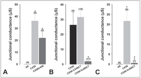
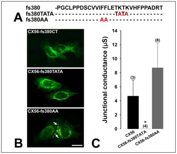
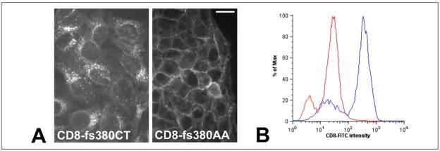
An increasing number of diseases have been mapped to genes coding for ion channel proteins, including the gap junction proteins, connexins. Here, we report on the identification of an amino acid sequence underlying the behavior of a non-functional mutant connexin46 (CX46) associated with congenital cataracts. The mutant protein, CX46fs380, is 31 amino acids longer than CX46 and contains 87 aberrant amino acids in its C terminus. When expressed in mammalian cells, the mutant CX46 was not found at gap junctional plaques, but it showed extensive co-localization with markers for ERGIC and Golgi. The severe reductions in function and formation of gap junctional plaques were transferred to other connexins by creating chimeras containing the last third (or more) of the aberrant C terminus of the CX46 mutant. This sequence also impaired trafficking of a CD8 chimera. Site-directed mutagenesis of a diphenylalanine restored appositional membrane localization and function. These results suggest a novel mechanism in which a mutation causes disease by generating a motif that leads to retention within the synthetic/secretory pathway.


Figures

FIGURE 1
FIGURE 1. Immunoreactive CX46fs380 localizes in the ERGIC and Golgi compartments
A, immunofluorescence of HeLa cells transiently transfected with wild-type CX46 (CX46), CX46 truncated after amino acid 379 (CX46tr380) or mutant CX46 (CX46fs380) after incubation with rabbit anti-CX46IL antibodies and Cy2-conjugated goat anti-rabbit IgG antibodies. Gap junctional plaques are indicated by arrows. B, confocal images of HeLa cells stably transfected with CX46fs380 under control conditions after double label immunofluorescence using anti-CX46fs380 antibodies (top panels) and a mouse monoclonal anti-ERGIC-53 (middle left panel) or anti-Golgi 58K protein (middle right panel) antibody; the overlap of the two signals is shown in the bottom panels. C, double label immunofluorescence of HeLa cells stably transfected with CX46fs380. Cells were incubated in the presence of cycloheximide for 16 h by which time negligible amounts of anti-CX46 immunoreactivity could be observed. Then, cells were incubated in the absence (CHX/Control) or presence of BFA (CHX/BFA). Immunoreactivity to anti-CX46fs380 is shown in green and that to anti-Golgi 58K protein is shown in red. The merged images are shown at the bottom; overlap of the signals appears yellow. Bar, 20 μm in A and C; 18 μm in B.
FIGURE 2
FIGURE 2. Connexin chimeras containing fs380CT exhibit impaired ability to form gap junctional plaques
HeLa cells transiently transfected with CX56 (CX56), CX56 truncated after amino acid 445 (CX56tr446) or CX56 chimeras containing the first 445 amino acids of CX56 and amino acids 380 – 435 of the C terminus of CX46 (CX56-CX46CT) or the aberrant sequence of CX46fs380 (CX56-fs380CT) were subjected to immunofluorescence using anti-CX56CT antibodies and Cy2-conjugated goat anti-rabbit IgG antibodies. Gap junctional plaques are indicated by arrows. Bar, 20 μm.
FIGURE 3
FIGURE 3. The CX56-fs380CT chimera is impaired in its ability to form functional gap junction channels
A–C, junctional conductances in pairs of Xenopus oocytes injected with cRNAs coding for CX56, CX56tr446, CX56-CX46CT, or CX56-fs380CT were determined by double whole cell voltage clamp. All oocytes were injected with antisense oligonucleotides (AS) to block endogenous Cx38 junctional currents. Data are presented as the mean ± S.E. The number of pairs is indicated in parentheses. *, p < 0.001 compared with AS-injected oocyte pairs; +, p < 0.001 when comparing CX56- or CX56-CX46CT-injected oocyte pairs with CX56-fs380CT-injected oocyte pairs.
FIGURE 4
FIGURE 4. A Cx32 chimera containing fs380CT is impaired in its capacity to form gap junctional plaques and induce junctional conductance
A, confocal images of HeLa cells stably transfected with Cx32-fs380CT after double label immunofluorescence using rabbit polyclonal anti-CX46fs380 antibodies (top panel) and a mouse monoclonal anti-PDI antibody (middle panel); the overlap of the two signals is shown on the bottom panel. B, junctional conductances in pairs of Xenopus oocytes injected with cRNAs coding for Cx32-CX46CT or Cx32-fs380CT were determined by double whole cell voltage clamp. Data are presented as the mean ± S.E. The number of pairs is indicated in parentheses. *, p < 0.001 compared with AS-injected oocyte pairs; #, p < 0.001 when comparing Cx32-CX46CT-injected oocyte pairs with Cx32-fs380CT-injected oocyte pairs. Bar, 10 μm.
FIGURE 5
FIGURE 5. The last 29 amino acids of the aberrant sequence of CX46fs380 are sufficient to induce loss of immunoreactivity at appositional membranes and loss of function
A, immunofluorescence of HeLa cells transiently transfected with CX56-fs380CT or chimeras containing the first one-third of fs380CT (CX56-fs380(–29)), the first two-thirds of the fs380CT sequence (CX56-fs380(1–58)) or the last one-third (CX56-fs380(59 – 87)) using rabbit anti-CX56CT antibodies and Cy2-conjugated goat anti-rabbit IgG antibodies. Gap junctional plaques are indicated by arrows. B and C, graphs showing the junctional conductances determined by double whole cell voltage clamp in pairs of Xenopus oocytes injected with CX56, CX56-fs380(1–58) or CX56-fs380(59 – 87) cRNAs. The data are presented as the mean ± S.E. The number of pairs is indicated in parentheses. *, p < 0.001 compared with AS-injected oocyte pairs; +, p < 0.001 when comparing CX56-injected oocyte pairs with CX56-fs380(59 – 87)-injected oocyte pairs. Bar, 20 μm.
FIGURE 6
FIGURE 6. A diphenylalanine in fs380CT is responsible for the aberrant localization and loss of function of CX56 chimeras
A, sequence of the last 29 amino acids of CX46fs380 (top line) and positions of the amino acids that were mutated to alanines (in red) to generate the fs380TATA and fs380AA mutant chimeras. B, immunofluorescence of HeLa cells transiently transfected with CX56-fs380CT or chimeras containing the aberrant sequence of CX46fs380 with two lysines (CX56-fs380TATA) or phenylalanines (CX56-fs380AA) substituted with alanines using rabbit anti-CX56CT antibodies and Cy2-conjugated goat anti-rabbit antibodies. Gap junctional plaques are indicated by arrows. C, graph showing the junctional conductances in pairs of Xenopus oocytes injected with CX56, CX56-fs380TATA, or CX56-fs380AA cRNAs. The data are presented as the mean ± S.E. of the number of pairs indicated in parentheses. +, p < 0.005 when comparing CX56-with CX56-fs380TATA-injected oocyte pairs. Bar, 20 μm.
FIGURE 7
FIGURE 7. Substitution of FF for AA restores membrane localization and function to a Cx32 chimera and CX46fs380
Immunofluorescence of HeLa cells transiently transfected with Cx32-fs380CT, Cx32-fs380AA (a chimera in which the diphenylalanines in fs380CT were mutated to dialanine), CX46fs380 or CX46fs380AA. Binding of rabbit polyclonal anti-Cx32 or anti-CX46IL antibodies was detected with Cy2-conjugated goat anti-rabbit IgG antibodies. Gap junctional plaques are indicated by arrows. Bar, 20 μm.
FIGURE 8
FIGURE 8. The diphenylalanine in fs380CT causes retention of a CD8 chimera
A, immunofluorescence of stably transfected HeLa cells showing the distribution of CD8 chimeras in which the cytoplasmic domain of CD8 was replaced with fs380CT (CD8-fs380CT) or with an fs380CT in which two alanines replaced the diphenylalanine (CD8-fs380AA). Localization of CD8 chimeras was detected using a FITC-conjugated antibody against the extracellular N terminus of CD8. B, graph illustrating the surface expression of CD8-fs380CT (red) and CD8-fs380AA (blue) measured by flow cytometry. Bar, 20 μm.

References

    1. Sáez JC, Berthoud VM, Branes MC, Martínez AD, Beyer EC. Physiol Rev. 2003;83:1359–1400. - PubMed
    1. Hübner CA, Jentsch TJ. Hum Mol Genet. 2002;11:2435–2445. - PubMed
    1. Reddy MA, Francis PJ, Berry V, Bhattacharya SS, Moore AT. Survey Ophthalmol. 2004;49:300–315. - PubMed
    1. Mackay D, Ionides A, Kibar Z, Rouleau G, Berry V, Moore A, Shiels A, Bhattacharya S. Am J Hum Genet. 1999;64:1357–1364. - PMC - PubMed
    1. Pal JD, Liu X, Mackay D, Shiels A, Berthoud VM, Beyer EC, Ebihara L. Am J Physiol Cell Physiol. 2000;279:C596–C602. - PubMed

Publication types

MeSH terms