. 2005 Oct 18;102(42):15122-7.
doi: 10.1073/pnas.0507346102. Epub 2005 Oct 4.

Requirement of myocardin-related transcription factor-B for remodeling of branchial arch arteries and smooth muscle differentiation

Affiliations

Requirement of myocardin-related transcription factor-B for remodeling of branchial arch arteries and smooth muscle differentiation

Jiyeon Oh et al. Proc Natl Acad Sci U S A. .

Abstract

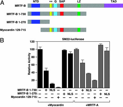
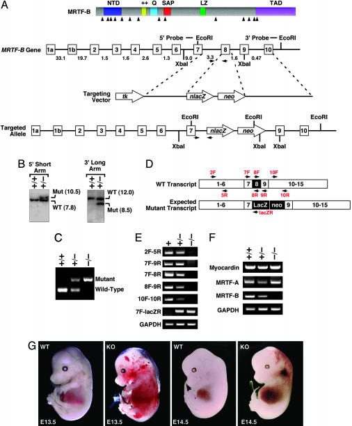
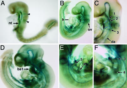
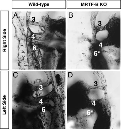
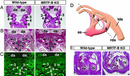
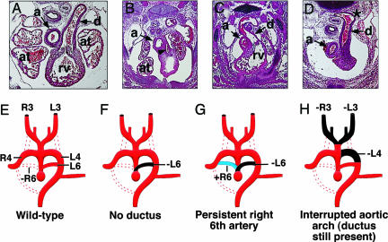
Myocardin and the myocardin-related transcription factors (MRTFs) A and B act as coactivators for serum response factor, which plays a key role in cardiovascular development. To determine the functions of MRTF-B in vivo, we generated MRTF-B mutant mice by targeted inactivation of the MRTF-B gene. We show that mice homozygous for an MRTF-B loss-of-function mutation die during mid-gestation from a spectrum of cardiovascular defects that includes abnormal patterning of the branchial arch arteries, double-outlet right ventricle, ventricular septal defects, and thin-walled myocardium. These abnormalities are accompanied by a failure in differentiation of smooth muscle cells within the branchial arch arteries, which are derived from the neural crest. The phenotype of MRTF-B mutant mice is distinct from that of mice lacking myocardin, revealing unique roles for these serum response factor coactivators in the development of different subsets of smooth muscle cells in vivo.


Figures

Fig. 1.
Fig. 1.
Transcriptional activities of MRTF-B mutant proteins. (A) The structures of wild-type and mutant MRTF-B and myocardin proteins are shown. The lacZ gene trap line (20) is predicted to generate a truncated MRTF-B protein containing residues 1–730. The targeted mutation we introduced into the MRTF-B gene is predicted to generate a truncated MRTF-B protein containing residues 1–270. A dominant negative mutant of myocardin is also shown. NTD, N-terminal domain; ++, basic domain; SAP, Scaffold attachment factor. Q, glutamine-rich domain; LZ, leucine zipper; TAD, transcription activation domain. (B) Wild type and mutant proteins were transfected into COS cells and assayed for their ability to activate an SM22-luciferase reporter. Truncated MRTF-B proteins containing the SV40 nuclear localization sequence (NLS) at the amino terminus were also tested. The MRTF-B 1–730 mutant functions as a dominant negative, whereas the 1–270 mutant lacks inhibitory activity.
Fig. 2.
Fig. 2.
Generation and analysis of MRTF-B knockout mice. (A) Gene targeting strategy. The mouse MRTF-B protein is schematized at the top. The targeting vector contained a 3.2-kb 5′ arm and a 4.7-kb 3′ arm and replaced a 0.5-kb region of the gene with a lacZ-neo cassette. Intron junctions within the coding region are shown by arrowheads beneath the schematized protein. Exons 1–10 are shown in boxes, and sizes of introns are indicated. Positions of 5′ and 3′ probes used for Southern analysis in B are indicated. Positions of PCR primers used for genotyping are shown by horizontal arrows. (B) Southern analysis. Genomic DNA from ES cell clones was isolated and analyzed by Southern blot with 5′ and 3′ probes after digestion with XbaI and EcoRI, respectively. (C) PCR analysis. Genomic DNA from E11.5 embryos was analyzed by PCR with primers shown in A.(D) Positions of primers used for RT-PCR. A schematic of the exons of the MRTF-B gene and positions of primers used for RT-PCR is shown. The expected mutation would contain a lacZ-neo cassette between exons 7 and 9 and would delete exon 8. (E) RT-PCR was performed by using RNA isolated from hearts of E10.5 embryos, and the primers shown in D. Genotypes of embryos are shown at the top. The truncated transcript generated from the mutant allele (2F-5R) was expressed at a much lower level than the WT MRTF-B transcript. (F) RT-PCR was performed by using RNA isolated from hearts of E10.5 embryos with primers specific for myocardin, MRTF-A, and MRTF-B. Transcripts for GAPDH were detected as a control for RNA loading and integrity. (G) Appearance of wild type and MRTF-B mutant embryos at E13.5 and E14.5.
Fig. 3.
Fig. 3.
LacZ staining of MRTF-B mutant embryos. Embryos were stained for lacZ expression. At E8.5, staining was apparent in the ventral neural tube and two distinct stripes (arrowheads) in the developing hindbrain (A). LacZ staining is observed in the otic vesicle, heart, dorsal aorta, and branchial arch arteries 1, 2, and 3 at E9.5 (B and C). At E10.5, lacZ expression becomes more widespread, with high level of expression in the first branchial arch (D). LacZ expression is also detected in the third and fourth arch arteries of MRTF-B mutant embryos as shown in the right and left lateral view (E and F). Branchial arch arteries are numbered. ba1, first branchial arch; nt, neural tube; ov, otic vesicle, h, heart; da, dorsal aorta.
Fig. 4.
Fig. 4.
Visualization of vasculature by India ink injection. India ink was injected into the beating hearts of wild-type (A and C) and MRTF-B–/– embryos (B and D) at E11.5. The right (A and B) and left (C and D) branchial arch arteries for each embryo are shown in lateral view and numbered. Note that both the right and left sixth arch arteries of mutant embryo are hypoplastic and prematurely regressed (*).
Fig. 5.
Fig. 5.
Branchial arch artery defects in MRTF-B mutant embryos. (AC) Wild-type and MRTF-B mutant embryos at E10.5 (A) and E11.5 (B and C). Hematoxylin/eosin sections show hypoplastic sixth arch arteries in mutant embryos. Abnormal communication between the fourth and sixth arteries at E10.5 is indicated (*). Histological sections stained for SM α-actin show smooth muscle differentiation within the wall of branchial arch arteries and dorsal aortae in the wild-type embryo. In contrast, SM α-actin expression is not detected in the branchial arch arteries of the mutant embryo, but expression is seen in the dorsal aorta and heart. Branchial arch arteries are numbered as 3, 4, and 6. da, dorsal aorta. (D) Schematic of branchial arch arteries. The plane of section in the embryos shown in AC is indicated by a dashed line. as, aortic sac; lda, left dorsal aorta; rda, right dorsal aorta. (E) Transverse section through the heart shows incomplete ventricular septation (arrowhead) and thin myocardial wall in the mutant embryo at E13.5. lv, left ventricle; rv, right ventricle.
Fig. 6.
Fig. 6.
Branchial arch artery defects in MRTF-B mutant embryos. (AD) Defects of the great vessels in E13.5 mutant embryos. In the transverse section of the wild-type embryo, the pulmonary trunk communicates with the descending aorta via the ductus arteriosus (A). Mutant embryos display hypoplastic pulmonary trunk (arrowhead) and no ductus arteriosus (B), persistent right-sided ductus arteriosus (C), and interrupted aortic arch (D). Note the abnormal shape and position of the descending aorta where the ductus arterious joins (*). a, aorta; at, atrium; d, ductus arteriosus; rv, right rentricle. (EH) Schematic diagrams of aortic arch defects seen in AD. Dotted lines represent normal regression. Black areas depict abnormal regression, whereas blue areas indicate abnormal persistence. (E) Patterning of the branchial arch arteries in the wild-type embryo with normal regression of the right sixth artery (-R6). (F) Absence of the ductus arteriosus resulting from abnormal regression of the left sixth artery (-L6). (G) Persistent right-sided ductus caused by abnormal regression of the left sixth artery (-L6) and abnormal persistence of the right sixth artery (+R6). (H) Interrupted aortic arch and loss of both carotid arteries, due to abnormal regression of the left fourth (-L4) and both right and left third arteries (-R3, -L3). R3, R4, and R6, right arch arteries 3, 4, and 6, respectively. L3, L4, and L6, left aortic arch arteries, respectively.

References

    1. Wang, D., Chang, P. S., Wang, Z., Sutherland, L., Richardson, J. A., Small, E., Krieg, P. A. & Olson, E. N. (2001) Cell 105, 851–862. - PubMed
    1. Wang, D.-Z., Li, S., Hockemeyer, D., Sutherland, L., Wang, Z., Schratt, G., Richardson, J. A., Nordheim, A. & Olson, E. N. (2002) Proc. Natl. Acad. Sci. USA 99, 14855–14860. - PMC - PubMed
    1. Wang, D.-Z. & Olson, E. N. (2004) Curr. Opin. Genet. Dev. 14, 558–566. - PubMed
    1. Cen, B., Selvaraj, A. & Prywes, R. (2004) J. Cell. Biochem. 93, 74–82. - PubMed
    1. Miano, J. M. (2003) J. Mol. Cell Cardiol. 35, 577–593. - PubMed

Publication types

MeSH terms