. 2005 Oct 18;102(42):15161-6.
doi: 10.1073/pnas.0507279102. Epub 2005 Oct 4.

Adaptive evolution of heparanase in hypoxia-tolerant Spalax: gene cloning and identification of a unique splice variant

Affiliations

Adaptive evolution of heparanase in hypoxia-tolerant Spalax: gene cloning and identification of a unique splice variant

Nicola J Nasser et al. Proc Natl Acad Sci U S A. .

Abstract

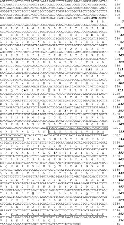
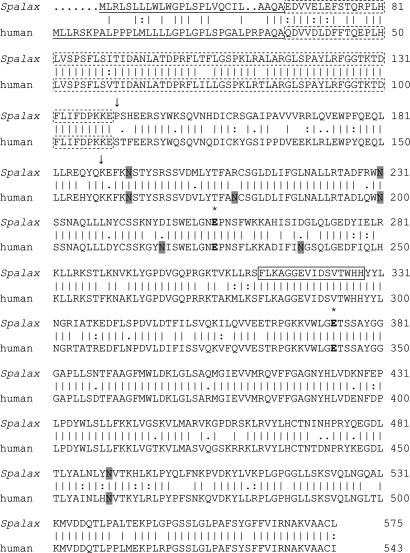
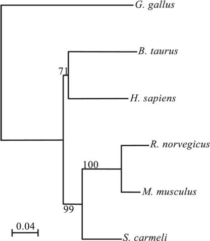
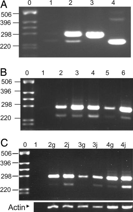
Heparan sulfate (HS) side chains of HS proteoglycans bind to and assemble extracellular matrix proteins and play important roles in cell-cell and cell-extracellular matrix interactions. HS chains bind a multitude of bioactive molecules and thereby function in the control of multiple normal and pathological processes. Enzymatic degradation of HS by heparanase, a mammalian endoglycosidase, affects the integrity and functional state of tissues and is involved in, among other processes, inflammation, angiogenesis, and cancer metastasis. Here, we report the cloning of heparanase from four Israeli species of the blind subterranean mole rat (Spalax ehrenbergi superspecies), 85% homologous to the human enzyme. Unlike its limited expression in human tissues, heparanase is highly expressed in diverse Spalax tissues. Moreover, we have identified a unique splice variant of the Spalax enzyme lacking 16 aa encoded by exon 7. This deletion resulted in a major defect in trafficking and processing of the heparanase protein, leading to a loss of its enzymatic activity. Interspecies variation was noted in the sequence and in the expression of the splice variant of the heparanase gene in blind mole rats living under different ecogeographical stresses, indicating a possible role in adaptation to stress in Spalax evolution.


Figures

Fig. 1.
Fig. 1.
Nucleotide and predicted amino acid sequences of Spalax heparanase. Nucleotide sequences are shown above the predicted amino acid sequences. Numbers on the right correspond to nucleotides (Roman) and amino acid residues (bold italic). The two initiation codons (ATG) and their corresponding methionine residues (M) are in bold. The three potential N-glycosylation sites are shaded. Arrowheads mark the two cleavage sites generating the two subunits and releasing the linker peptide residing in between. The nucleotide and amino acid sequences lacking in splice 7 are boxed. The hydrophobic potential membrane-spanning domain of 19 aa is underlined.
Fig. 2.
Fig. 2.
Comparison of Spalax and human heparanase amino acid sequences. Vertical lines denote conserved amino acids, and double or single dots mark similar amino acids (Wisconsin Package, Version 103). The putative two catalytic Glu residues, the proton donor and the nucleophile, are marked in bold with * above. The potential N-glycosylation sites are shaded. The 8-kDa subunit is marked with a dotted box. The cleavage sites generating the mature enzyme are marked by arrows, and amino acids between the two arrows denote the linker sequence. The sequence boxed with a continuous line denotes the amino acids lacking in splice 7 of the Spalax heparanase.
Fig. 3.
Fig. 3.
Heparanase similarity tree: an amino acid-based tree using the Kimura distances. The bar represents substitutions per amino acid. The numbers in the junctions are bootstrapping (in percentage) based on 1,000 replications. Alignment of the Spalax amino acids sequence with that of the rat, mouse, human, bovine, and chicken shows 86.7%, 88.6%, 85%, 83.7%, and 67.2% identity, respectively.
Fig. 4.
Fig. 4.
Expression of heparanase in different Spalax tissues. Semiquantitive RT-PCR using Spalax-specific primers located around the heparanase cDNA region encoded by exon 7. Bands of 288 bp represent the wild-type enzyme, and those of 240 bp represent its splice 7 form. (A) Lane 1, reaction mixture alone; lane 2, cDNA of kidney; lanes 3 and 4, plasmids containing the cDNA sequence of the wild-type Spalax heparanase and the splice 7 variant, respectively. (B) Lane 1, reaction mixture alone; lanes 2–6, cDNAs of S. judaei kidney, liver, heart, brain, and eye, respectively. (C) Comparison of heparanase expression of S. galili and S. judaei (g or j added to the lane number, respectively). Lane 1, reaction mixture alone; lanes 2–4, cDNAs from kidney, brain, and liver, respectively. The same cDNA preparations were subjected to RT-PCR using primers specific for Spalax β-actin to control for equal loading. Note the higher expression of splice 7 in S. judaei. DNA ladder lanes are marked by 0. Shown to the left of the DNA ladder are the corresponding number of base pairs.
Fig. 5.
Fig. 5.
Expression, glycosylation, secretion, and enzymatic activity of splice 7 vs. Spalax heparanases. (A–C) Western blots using anti-heparanase antibodies 1453 in A and C and 810 in B. (A) Lysates of 293HEK cells transfected with mock (lane 1), human (lane 2), or Spalax (lane 3) heparanases. (B) 293HEK cells transfected with mock (lane 1), human (lanes 2 and 4), or Spalax (lanes 3 and 5) heparanases were preincubated without (lanes 2 and 3) or with (lanes 4 and 5) tunicamycin. Cell lysates were subjected to SDS/PAGE and Western blotting, as described in Materials and Methods. Note that the molecular weight difference between the human (lane 2) and Spalax (lane 3) heparanases is abolished after treatment with tunicamycin (lanes 4 and 5). (C) Comparison of Spalax wild-type and splice 7 heparanase processing, secretion, and heparin binding. The first blot shows lysates, and the second blot shows conditioned medium of cells transfected with mock (lane 1), Spalax wild-type (lane 2), and splice 7 (lane 3) heparanases. Note the lack of processing (first blot) and secretion (second blot) of splice 7. The third and fourth blots show heparin-binding capacity. Lysates of 293HEK cells transfected with mock (lanes 1), Spalax wild-type (lane 2), or splice 7 (lane 3) heparanases were incubated with Fractogel (third blot), as a positive control, or with heparin beads (fourth blot). Proteins remaining bound to the resin and beads after washing were subjected to Western blot analysis using anti-heparanase antibodies, as described in Materials and Methods. Both wild-type and splice 7 Spalax heparanases bind to the heparin beads. (D) Heparanase enzymatic activity. Lysates of cell stably transfected with pcDNA3 vectors containing Spalax wild-type (♦) or splice 7 (□) heparanases vs. mock, insert-free plasmid alone (▪) were incubated (4 h, 37°C, pH 6.0) with 35S-labeled ECM. Labeled degradation fragments released into the incubation medium were analyzed by gel filtration on Sepharose 6B. Peak I (fractions 1–10), representing nearly intact HS proteoglycans, was noticed in the mock (▪) and splice 7 (□) transfected cells. Peak II (fractions 20–30), representing HS degradation products, was obtained in cells transfected with the wild-type Spalax heparanase (♦).

References

    1. Kjellen, L. & Lindahl, U. (1991) Annu. Rev. Biochem. 60, 443-475. - PubMed
    1. Bernfield, M., Gotte, M., Park, P. W., Reizes, O., Fitzgerald, M. L., Lincecum, J. & Zako, M. (1999) Annu. Rev. Biochem. 68, 729-777. - PubMed
    1. Iozzo, R. V. (1998) Annu. Rev. Biochem. 67, 609-652. - PubMed
    1. Vlodavsky, I., Bar-Shavit, R., Korner, G. & Fuks, Z. (1993) in Basement Membranes: Cellular and Molecular Aspects, eds. Rohrbach, D. H. & Timpl, R. (Academic, Orlando, FL), pp. 327-343.
    1. Vlodavsky, I., Miao, H.-Q., Medalion, B., Danagher, P. & Ron, D. (1996) Cancer Metastasis Rev. 15, 177-186. - PubMed

Publication types

MeSH terms

LinkOut - more resources