. 2005;7(5):R1034-45.
doi: 10.1186/ar1779. Epub 2005 Jun 23.

Protective effect of vasoactive intestinal peptide on bone destruction in the collagen-induced arthritis model of rheumatoid arthritis

Affiliations

Protective effect of vasoactive intestinal peptide on bone destruction in the collagen-induced arthritis model of rheumatoid arthritis

Yasmina Juarranz et al. Arthritis Res Ther. 2005.

Abstract

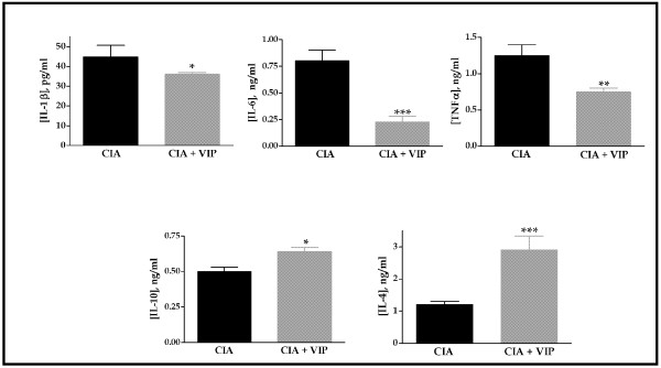
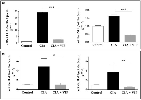
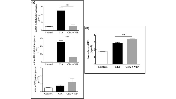
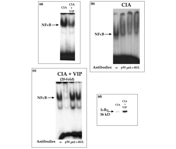
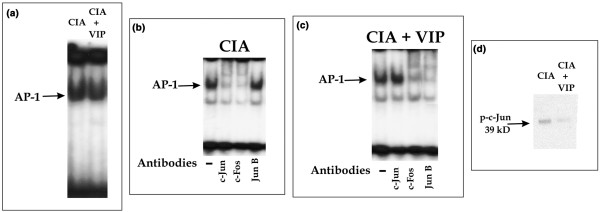
Rheumatoid arthritis (RA) is an autoimmune disease of unknown etiology, characterized by the presence of inflammatory synovitis accompanied by destruction of joint cartilage and bone. Treatment with vasoactive intestinal peptide (VIP) prevents experimental arthritis in animal models by downregulation of both autoimmune and inflammatory components of the disease. The aim of this study was to characterize the protective effect of VIP on bone erosion in collagen-induced arthritis (CIA) in mice. We have studied the expression of different mediators implicated in bone homeostasis, such as inducible nitric oxide synthase (iNOS), cyclooxygenase-2 (COX-2), receptor activator of nuclear factor-kappaB (RANK), receptor activator of nuclear factor-kappaB ligand (RANKL), osteoprotegerin (OPG), IL-1, IL-4, IL-6, IL-10, IL-11 and IL-17. Circulating cytokine levels were assessed by ELISA and the local expression of mediators were determined by RT-PCR in mRNA extracts from joints. VIP treatment resulted in decreased levels of circulating IL-6, IL-1beta and TNFalpha, and increased levels of IL-4 and IL-10. CIA-mice treated with VIP presented a decrease in mRNA expression of IL-17, IL-11 in the joints. The ratio of RANKL to OPG decreased drastically in the joint after VIP treatment, which correlated with an increase in levels of circulating OPG in CIA mice treated with VIP. In addition, VIP treatment decreased the expression of mRNA for RANK, iNOS and COX-2. To investigate the molecular mechanisms involved, we tested the activity of NFkappaB and AP-1, two transcriptional factors closely related to joint erosion, by EMSA in synovial cells from CIA mice. VIP treatment in vivo was able to affect the transcriptional activity of both factors. Our data indicate that VIP is a viable candidate for the development of treatments for RA.


Figures

Figure 1
Figure 1
Cytokine circulating levels in mice at the end of treatment in the collagen-induced arthritis (CIA) model. IL-1β, tumor necrosis factor (TNF)α, IL-6, Il-10 or IL-4 were measured (mean ± SEM) by ELISA in arthritic animals and the same animals treated with VIP. On day 10 of VIP treatment, differences between the arthritic group and the CIA group treated with vasoactive intestinal peptide (VIP) were statistically significant (*p < 0.05, **p < 0.01, ***p < 0.001). Results are the mean ± SEM of two separate experiments with 10 animals per group.
Figure 2
Figure 2
mRNA expression of inflammatory mediators and cytokines related to bone destruction. (a) Expression of mRNA for cyclooxygenase-2 (COX-2) and inducible nitric oxide synthase (iNOS) in the hind paws was measured by quantitative real-time PCR and corrected by mRNA expression for β-actin in each sample (see Materials and methods). (b) Expression of mRNA for IL-11 and IL-17 in the hind paws was measured by quantitative real-time PCR and corrected by mRNA expression for β-actin in each sample (see Materials and methods). On day 10 of vasoactive intestinal peptide (VIP) treatment, differences between the arthritic group and the CIA group treated with VIP were statistically significant (*p < 0.05, **p < 0.01, ***p < 0.001). Results are the mean ± SEM of two separate experiments with 10 animals per group.
Figure 3
Figure 3
Vasoactive intestinal peptide (VIP) modulates the pattern of expression of the RANK/RANKL/OPG system in joints from mice with collagen-induced arthritis (CIA). (a) Expression of mRNA for receptor activator of nuclear factor-κB (RANK), receptor activator of nuclear factor-κB ligand (RANKL) or osteoprotegerin (OPG) in the hind paws was measured by quantitative real time PCR and corrected by mRNA expression for β-actin in each sample (see Materials and methods). (b) Serum levels of OPG in control, CIA or VIP-treated CIA mice were determined by ELISA. On day 10 of VIP treatment, differences between the arthritic group and the CIA group treated with VIP were statistically significant (**p < 0.01, ***p < 0.001). Results are the mean ± SEM of two independent experiments with 10 animals per group
Figure 4
Figure 4
Effect of vasoactive intestinal peptide (VIP) on NFκB binding and IκB degradation in synovial cells from mice with collagen-induced arthritis (CIA). (a) EMSA results from nuclear extracts of synovial cells from CIA or VIP-treated CIA mice, using a radiolabeled oligonucleotide containing the NFκB consensus binding site. (b) Supershift assay on nuclear extracts of CIA mice using anti-p50, anti-p65 or anti-cRel. (c) Supershift assay (20-fold amplified) on nuclear extracts of VIP-treated CIA mice using anti-p50, anti-p65 or anti-cRel. (d) Western blot analysis showing immunoreactive IκBα (36 kDa) in cytoplasmic fractions of synovial cells from CIA and VIP-treated CIA mice. A representative experiment of three is shown.
Figure 5
Figure 5
AP-1 binding and c-Jun activation in synovial cells from mice with collagen-induced arthritis (CIA) after vasoactive intestinal peptide (VIP) treatment. (a) EMSA results from nuclear extracts of synovial cells from CIA or VIP-treated CIA mice, using a radiolabeled oligonucleotide containing the AP-1 consensus binding site. (b) Supershift assay on nuclear extracts of CIA mice using anti-c-Jun, anti-c-Fos or anti-Jun B. (c) Supershift assay on nuclear extracts of VIP-treated CIA mice using anti-c-Jun, anti-c-Fos or anti-Jun B. (d) Western blot analysis showing immunoreactive phosphorylated c-Jun (39 kDa) in cytoplasmic fractions of synovial cells from CIA and VIP-treated CIA mice. A representative experiment of three is shown.

Similar articles

Cited by

References

    1. Gravallese EM. Bone destruction in arthritis. Ann Rheum Dis. 2002;61:ii84–ii86. - PMC - PubMed
    1. Takayanagi H, Iizuka H, Juji T, Nakagawa T, Yamamoto A, Myazaki T, Koshihara Y, Oda H, Nakamura K, Tanaka S. Involvement of receptor activator of nuclear factor kB ligand/osteoclast differentiation factor in osteoclastogenesis from synoviocytes in rheumatoid arthritis. Arthritis Rheum. 2000;43:259–269. doi: 10.1002/1529-0131(200002)43:2<259::AID-ANR4>3.0.CO;2-W. - DOI - PubMed
    1. Hofbauer LC, Heulfelder AE. The role of osteoprotegerin and receptor activator of nuclear factor kappaB ligand in the pathogenesis and treatment of rheumatoid arthritis. Arthritis Rheum. 2001;44:253–259. doi: 10.1002/1529-0131(200102)44:2<253::AID-ANR41>3.0.CO;2-S. - DOI - PubMed
    1. Nakashima T, Wada T, Penninger JM. RANKL and RANK as novel therapeutic targets for arthritis. Curr Opin Rheumatol. 2003;15:280–287. doi: 10.1097/00002281-200305000-00016. - DOI - PubMed
    1. O'Gradaigh D, Ireland D, Bord S, Compston JE. Joint erosion in rheumatoid arthritis: interactions between tumor necrosis factor α, interleukin 1, and receptor activator of nuclear factor kB ligand (RANKL) regulate osteoclasts. Ann Rheum Dis. 2004;63:354–359. doi: 10.1136/ard.2003.008458. - DOI - PMC - PubMed

Publication types

MeSH terms