

Review
doi: 10.1186/gb-2005-6-10-233.
Epub 2005 Sep 29.
Global approaches to understanding ubiquitination
Affiliations
- PMID: 16207362
- PMCID: PMC1257457
- DOI: 10.1186/gb-2005-6-10-233
Item in Clipboard
Review
Global approaches to understanding ubiquitination
Genome Biol.
2005.
Abstract
Ubiquitination--the linkage of one or more molecules of the protein ubiquitin to another protein--regulates a wide range of biological processes in all eukaryotes. We review the proteome-wide strategies that are being used to study aspects of ubiquitin biology, including substrates, components of the proteasome and ubiquitin ligases, and deubiquitination.
Figures
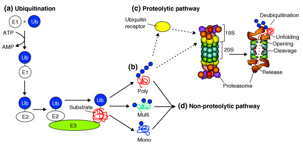
The ubiquitin proteasome system. (a) Ubiquitin is activated by a ubiquitin-activating enzyme (E1) and transferred onto substrate proteins by ubiquitin-conjugating enzymes (E2) and ubiquitin ligases (E3), resulting in (b) either attachment of a single ubiquitin molecule (mono-ubiquitination), attachment of multiple ubiquitin units to several substrate lysine residues on the same protein (multi-ubiquitination) or synthesis of ubiquitin chains (poly-ubiquitination). (c) Many poly-ubiquitinated proteins are subsequently degraded by the 26S proteasome, which consists of the catalytic 20S complex and the regulatory 19S particles. Degradation substrates are either delivered to the proteasome by soluble ubiquitin receptors or recognized by the intrinsic ubiquitin-binding activity of the 19S particle. At the 19S proteasome the ubiquitin chain is disassembled, and the substrate is unfolded before it can enter the cavity of the 20S subunit where proteolysis takes place. Finally, proteolytic fragments exit the proteasome in a poorly understood way. (d) Ubiquitination can also directly regulate protein function in a proteolysis-independent manner, via mono-, multi- or poly-ubiquitinated proteins.
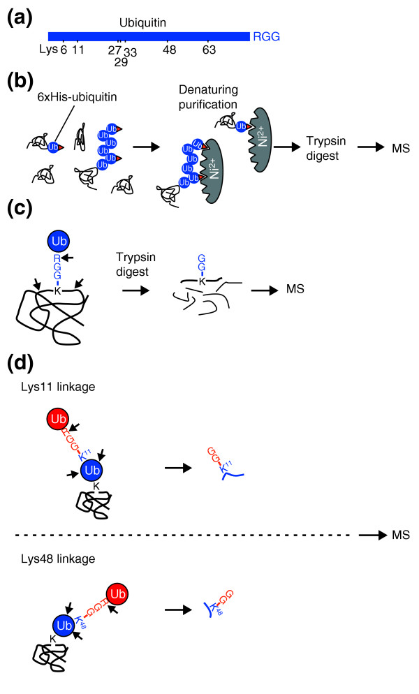
Global strategies that use mass spectrometry (MS) to study ubiquitination. (a) Diagram of the lysine residues in ubiquitin; the carboxy-terminal Arg-Gly-Gly (RGG) motif is also indicated. (b) In ubiquitin profiling, 6×His-tagged ubiquitin expressed in cells is conjugated to substrate proteins, and this facilitates purification of ubiquitinated proteins under denaturing conditions by Ni-chelate chromatography, in which histidine-tagged proteins bind specifically to immobilized Ni2+ ions. Purified ubiquitinated proteins are digested with trypsin and the resulting peptides are analyzed by mass spectrometry to identify the proteins present in the sample. (c) Precise ubiquitination sites can be determined by mass spectrometry because of a characteristic mass shift caused by diglycine that is retained on ubiquitinated lysine residues within peptides after trypsin digestion. (d) A similar strategy allows differentiation between the various types of ubiquitin chain linkage that can lead to diverse ubiquitin-chain topologies. Depending on the lysine residue in ubiquitin that was used for the ubiquitin-ubiquitin linkage, different linkage-specific signature peptides with characteristic masses are produced by trypsin digestion. These signature peptides can be detected and distinguished by mass spectrometry.
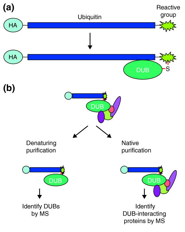
Activity-based profiling of deubiquitinating enzymes and interacting proteins. (a) Ubiquitin fused to an amino-terminal epitope tag (for example hemagglutinin, HA) and a carboxy-terminal reactive group forms a covalent conjugate with deubiquitinating enzymes (DUBs; for details of the generation of these ubiquitin probes, see [57]). (b) The DUB-ubiquitin conjugates can be immunopurified using the HA epitope. Immunopurification under native conditions allows identification of DUBs and their interacting proteins by mass spectrometry (MS). The immunopurified fractions can be further separated by gel electrophoresis, and DUB-ubiquitin conjugates can be detected by anti-HA immunoblotting. Proteins corresponding to HA-reactive bands can be eluted from silver-stained gels (not shown) and the DUBs can be identified by mass spectrometry.
References
Publication types
MeSH terms
Substances
Grants and funding
LinkOut - more resources
Full Text Sources
Molecular Biology Databases
