

Malignant schwannoma of the upper mediastinum originating from the vagus nerve
- PMID: 16207383
- PMCID: PMC1276821
- DOI: 10.1186/1477-7819-3-65
Malignant schwannoma of the upper mediastinum originating from the vagus nerve
Abstract
Background: Malignant schwannoma of the upper mediastinum originating from the vagus nerve is extremely rare.
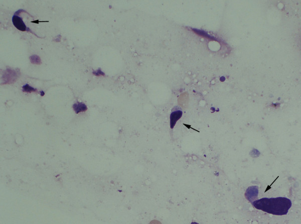
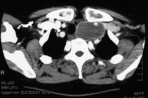
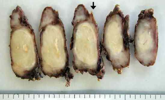
Case presentation: A 46-year-old female was admitted for a left cervical mass which was associated with both hoarseness and Horner's syndrome. Chest computed tomography showed a mass extending from the left upper mediastinum to the left supraclavicular area. A fine needle aspiration cytological examination suggested primary lung cancer stage IIIB large cell carcinoma. After administering induction chemo-radiotherapy, a complete surgical resection was performed. The tumor was found to involve both the left vagus nerve and the left sympathetic nerve. Histological examination of the resected specimen revealed the tumor to be malignant schwannoma.
Conclusion: Despite incorrect preoperative diagnosis, the multimodality treatment administered in this case, including induction chemo-radiotherapy and surgery, proved to be effective.
Figures

References
-
- Davis RD, Oldham HN, Sabiston DC. Primary cysts and neoplasms of the mediastinum: recent changes in clinical presentation, methods of diagnosis, management, and results. Ann Thorac Surg. 1987;44:229–237. - PubMed
-
- Travis WD, Colby TV, Corrin B. World Health Organization International Histological Classification of Tumors. Histological typing of Lung and Pleural Tumors, 3 rd ed. Berlin: Springer Verlag; 1999.
LinkOut - more resources
Full Text Sources
