. 2005 Dec;16(12):5891-900.
doi: 10.1091/mbc.e05-07-0660. Epub 2005 Oct 5.

BAG-2 acts as an inhibitor of the chaperone-associated ubiquitin ligase CHIP

Affiliations

BAG-2 acts as an inhibitor of the chaperone-associated ubiquitin ligase CHIP

Verena Arndt et al. Mol Biol Cell. 2005 Dec.

Abstract

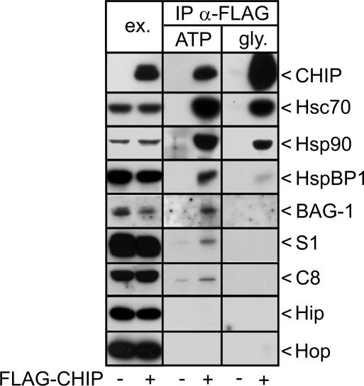
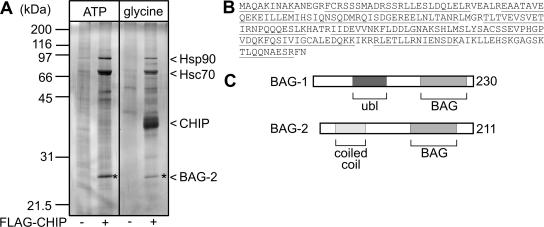
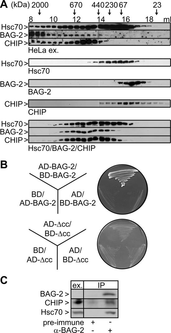
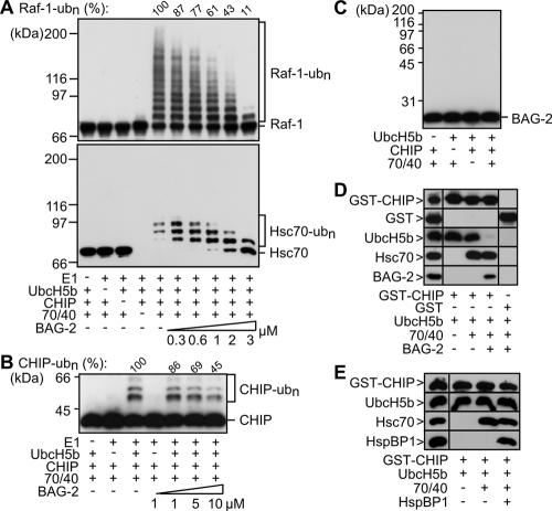
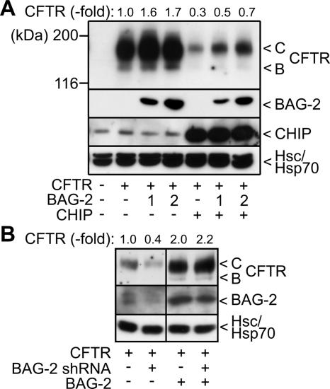
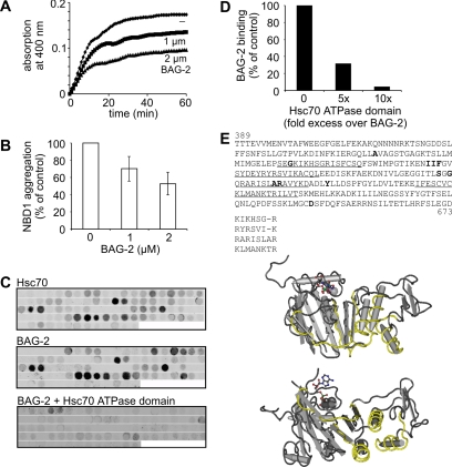
Cellular protein quality control involves a close interplay between molecular chaperones and the ubiquitin/proteasome system. We recently identified a degradation pathway, on which the chaperone Hsc70 delivers chaperone clients, such as misfolded forms of the cystic fibrosis transmembrane conductance regulator (CFTR), to the proteasome. The cochaperone CHIP is of central importance on this pathway, because it acts as a chaperone-associated ubiquitin ligase. CHIP mediates the attachment of a ubiquitin chain to a chaperone-presented client protein and thereby stimulates its proteasomal degradation. To gain further insight into the function of CHIP we isolated CHIP-containing protein complexes from human HeLa cells and analyzed their composition by peptide mass fingerprinting. We identified the Hsc70 cochaperone BAG-2 as a main component of CHIP complexes. BAG-2 inhibits the ubiquitin ligase activity of CHIP by abrogating the CHIP/E2 cooperation and stimulates the chaperone-assisted maturation of CFTR. The activity of BAG-2 resembles that of the previously characterized Hsc70 cochaperone and CHIP inhibitor HspBP1. The presented data therefore establish multiple mechanisms to control the destructive activity of the CHIP ubiquitin ligase in human cells.


Figures

Figure 1.
Figure 1.
Protein composition of CHIP-containing protein complexes isolated from HeLa cells. HeLa cells were transfected with pCMVTag2b-chip, which encodes a FLAG-tagged form of CHIP (+ FLAG-CHIP), or with an equal amount of a control plasmid (-). CHIP-containing protein complexes were precipitated with an immobilized anti-FLAG antibody (IP α-FLAG). Purified immunocomplexes were treated with ATP (ATP), followed by addition of glycine, pH 3.5, to release CHIP and remaining associated proteins from the antibody (gly.). CHIP immunocomplexes were separated by SDS-PAGE and analyzed for the presence of binding partners by immunoblotting with specific antibodies. To monitor expression levels, 40 μg of protein extracts were loaded (ex.).
Figure 2.
Figure 2.
The cochaperone BAG-2 is a major component of CHIP-containing protein complexes. (A) Analysis of isolated CHIP immmunocomplexes by SDS-PAGE and silver-staining. Hsp90, Hsc70, and CHIP were identified according to their molecular masses. The star denotes a prominent polypeptide of ∼27 kDa. The corresponding polypeptide was identified as the cochaperone BAG-2 by peptide mass fingerprinting. (B) Complete sequence of human BAG-2. Polypeptides of BAG-2 identified by mass spectrometry are underlined. (C) Schematic presentation of the domain structure of BAG-1 and BAG-2. Both cochaperones possess a BAG domain at their carboxyl termini, which mediates Hsc70 binding and regulation. In addition, BAG-1 carries a ubiquitin-like domain (ubl), whereas BAG-2 possesses a coiled-coil domain near the amino terminus.
Figure 3.
Figure 3.
BAG-2 forms a complex with Hsc70 and CHIP. (A) HeLa cells were transfected with pCMVTag2b-bag-2, which encodes a FLAG-tagged form of BAG-2 (+ FLAG-BAG-2), or with an equal amount of a control plasmid (-). BAG-2-containing protein complexes were immunoprecipitated with an immobilized anti-FLAG antibody (IP α-FLAG) and analyzed for the presence of Hsc70 and CHIP by immunoblotting. The chaperone and CHIP were detected in ATP-eluates of the isolated complexes. To monitor expression levels, 40 μg of protein extracts was loaded (ex.). Twenty percent of each IP fraction was loaded. (B) Binding of BAG-2 to CHIP is mediated by Hsc70. Purified GST-tagged BAG-2 was immobilized and incubated with Hsc70 and CHIP as indicated. Proteins retained on the affinity resin were detected using specific antibodies. As negative control, proteins were incubated with immobilized GST. The left panel shows 5% of the input. (C) Schematic presentation of the BAG-2/Hsc70/CHIP complex. BAG-2 binds to the ATPase domain of Hsc70 (A) utilizing its BAG domain. At the same time CHIP associates with the carboxy terminus of the chaperone (C) via its TPR domain. P denotes the peptide-binding pocket of Hsc70. (D) BAG-2 stimulates the association of Hsc70 with CHIP. GST-CHIP was immobilized and incubated with Hsc70 and BAG-2 as indicated. BAG-2 was added at 1-, 2-, and 4-fold the concentration of CHIP. Bound proteins were detected after immunoblotting using specific antibodies. Bound Hsc70 was quantified, and values are presented as percentage of control in the absence of BAG-2, which was set to 100%. (E) Overexpression of BAG-2 increases the amount of Hsc70 detectable in CHIP-containing protein complexes. HeLa cells were transfected with pCMVTag2b-chip (+ FLAG-CHIP) and pcDNA3.1-bag-2 (+ BAG-2) as indicated. The total amount of added DNA was kept constant by addition of pcDNA3.1. CHIP-containing protein complexes were isolated using an immobilized anti-FLAG antibody (IP α-FLAG). Immunoprecipitated CHIP was detected in glycine eluates using a specific antibody. BAG-2 and Hsc70 were detected in ATP eluates of the isolated CHIP complexes by immunoblotting.
Figure 4.
Figure 4.
BAG-2 cofractionates with Hsc70 and CHIP in a HeLa cell extract and forms homodimers. (A) Analysis of chaperone/cochaperone complexes by gel filtration chromatography. Top, fractionation of a HeLa cell extract on a Superose 6 column. Proteins were detected by specific antibodies as indicated on the left. The elution behavior of proteins of defined molecular mass is indicated at the top. Bottom, gel filtration analysis of purified components and Hsc70/BAG-2/CHIP complexes formed in vitro. (B) Plasmid constructs encoding BAG-2 or a deletion fragment of BAG-2 (ΔCC) fused to the GAL4-transcription activation domain (AD-BAG-2/-ΔCC) or the GAL4-DNA-binding domain (BD-BAG-2/-ΔCC) and empty vectors (AD and BD) were transformed into yeast and screened for potential interactions in a yeast two-hybrid assay. Dimerization of BAG-2 resulted in growth on minimal medium lacking histidine, leucine, and tryptophan. (C) Coimmunoprecipitation of endogenous BAG-2, CHIP, and Hsc70. HeLa cell extracts were subjected to immunoprecipitation with an anti-BAG-2 antibody. Preimmune serum was used in control reactions. CHIP and Hsc70 were detected in ATP eluates of the isolated immunocomplexes. BAG-2 was eluted with synthetic peptide. Ex. corresponds to 60 μg of HeLa cell extract. Under the chosen conditions endogenous BAG-2 is not detectable in the extract.
Figure 5.
Figure 5.
BAG-2 inhibits the ubiquitin ligase activity of CHIP when complexed with Hsc70 by abrogating CHIP/E2 cooperation. (A) Ubiquitylation of Raf-1 and Hsc70 was analyzed in vitro in the presence of purified CHIP (1 μM), the ubiquitin-activating enzyme E1, UbcH5b, Hsc70/Hsp40 (70/40), and increasing concentrations of BAG-2, ranging from 0.3 to 3 μM, as indicated. Raf-1, Hsc70, and ubiquitylated forms of both proteins (ub(n)-Raf-1, ub(n)-Hsc70) were detected using specific antibodies. Ubiquitylated Raf-1 was quantified, and obtained values are given as percentage of the amount detected in a control reaction in the absence of BAG-2. Notably, Hsc70 that carried more than three ubiquitin moieties was not recovered under the experimental conditions. (B) Autoubiquitylation of CHIP was investigated in vitro in the presence of purified CHIP (1 μM), the ubiquitin-activating enzyme E1, UbcH5b, Hsc70/Hsp40 (70/40), and increasing concentrations of BAG-2, ranging from 1 to 10 μM. Ubiquitylated forms of CHIP (ub(n)-CHIP) were detected by immunoblotting using specific antibodies and their amount was quantified. (C) BAG-2 is not recognized as a substrate by the CHIP ubiquitin ligase. In vitro ubiquitylation reactions were performed as in A except that the reactions contained purified BAG-2 instead of Raf-1. BAG-2 was detected in immunoblots using a specific antibody. In the presented experiment, CHIP-mediated ubiquitylation of Hsc70 was reduced to 26% in the presence of BAG-2 compared with a control reaction without BAG-2 which was set to 100%. (D) GST-CHIP was immobilized and incubated with UbcH5b, Hsc70/Hsp40 (70/40), and BAG-2 as indicated. Bound proteins were detected after immunoblotting using specific antibodies. As a control proteins were incubated with immobilized GST (right panel). The panel on the left shows 10% of the input. (E) In contrast to BAG-2, HspBP1 does not interfere with the CHIP/E2 interaction. UbcH5b binding to immobilized CHIP was analyzed as described in D in the presence of HspBP1 instead of BAG-2.
Figure 6.
Figure 6.
BAG-2 inhibits the chaperone-assisted degradation of CFTR and is essential for CFTR maturation. (A) HEK293 cells were transiently transfected with CFTR-, BAG-2-, and CHIP-encoding plasmids in the indicated combinations. Amounts of pcDNA3.1-bag-2 were 1- and 2-fold of the amount of pcDNA3.1-chip when indicated. The ER-localized, core-glycosylated B-form (B) and the completely glycosylated, plasma membrane-localized C-form of CFTR (C) were detected by immunoblotting. Likewise, BAG-2 and CHIP were detected with specific antibodies. Hsc70 and Hsp70 served as loading control. Each lane represents 60 μg of cellular extract. CFTR levels were quantified from four independent experiments and mean values are presented as fold change compared with the control reaction, which was set to 1. (B) HEK293 cells were transiently transfected with CFTR-, BAG-2 shRNA-, and BAG-2-expressing plasmids as indicated. Cell extracts, 60 μg, were separated by SDS-PAGE. Protein levels were analyzed by immunoblotting using specific antibodies. Hsc70/Hsp70 served as loading control. Samples that did not express BAG-2 shRNA received the pSUPER plasmid without an insert.
Figure 7.
Figure 7.
BAG-2 inhibits the aggregation of the ΔF508 NBD1 domain of CFTR by binding hydrophobic and positively charged segments in NBD1. (A) Aggregation of denatured NBD1 was followed over time at 37°C by turbidity measurement at 400 nm. NBD1, 2 μM, was incubated either alone or in the presence of purified BAG-2 at the indicated concentrations. (B) Data obtained in A are presented as the relative amount of aggregated NBD1 domain after a 60-min incubation period. The amount of aggregated NBD1 in the control reaction was set as 100%. Values represent the mean of 14 independent experiments plus SD. (C) Fluorophor-labeled Hsc70 and BAG-2 were incubated with peptide scans covering NBD1, and retained protein was visualized with a fluorescence scanner. When indicated, binding of labeled BAG-2 to NBD1 peptides was performed in the presence of the Hsc70 ATPase domain at a 10-fold molar excess over the cochaperone. (D) Binding of fluorophor-labeled BAG-2 to aa 589-601 of the NBD1 domain either alone or in the presence of a 5- or 10-fold molar excess of the Hsc70 ATPase domain was quantified. The amount of BAG-2 bound to aa 589-601 in the absence of the ATPase domain was set to 100%. (E) Primary structure of NBD1 of human CFTR. Amino acids shown to be mutated in cystic fibrosis patients are highlighted in bold. Sequences recognized by BAG-2 and Hsc70 are underlined. Comparison of identified sequences reveals the presence of an interaction motif that is characterized by a core of hydrophobic amino acids flanked by positively charged amino acids. Identified binding regions are shown in yellow in the resolved NBD1 structure (1XMJ_A of the NCBI structure database).

Similar articles

Cited by

References

    1. Alberti, S., Demand, J., Esser, C., Emmerich, N., Schild, H., and Höhfeld, J. (2002). Ubiquitylation of BAG-1 suggests a novel regulatory mechanism during the sorting of chaperone substrates to the proteasome. J. Biol. Chem. 277, 45920-45927. - PubMed
    1. Alberti, S., Böhse, K., Arndt, V., Schmitz, A., and Höhfeld, J. (2004). The co-chaperone HspBP1 inhibits the CHIP ubiquitin ligase and stimulates the maturation of the cystic fibrosis transmembrane conductance regulator. Mol. Biol. Cell 15, 4003-4010. - PMC - PubMed
    1. Ballinger, C. A., Connell, P., Wu, Y., Hu, Z., Thompson, L. J., Yin, L.Y., and Patterson, C. (1999). Identification of CHIP, a novel tetratricopeptide repeat-containing protein that interacts with heat shock proteins and negatively regulates chaperone functions. Mol. Cell. Biol. 19, 4535-4545. - PMC - PubMed
    1. Cardozo, C. P., Michaud, C., Ost, M. C., Fliss, A. E., Yang, E., Patterson, C., Hall, S. J., and Caplan, A. J. (2003). C-terminal Hsp-interacting protein slows androgen receptor synthesis and reduces its rate of degradation. Arch. Biochem. Biophys. 410, 134-140. - PubMed
    1. Connell, P., Ballinger, C. A., Jiang, J., Wu, Y., Thompson, L. J., Höhfeld, J., and Patterson, C. (2001). The co-chaperone CHIP regulates protein triage decisions mediated by heat-shock proteins. Nat. Cell Biol. 3, 93-96. - PubMed

Publication types

MeSH terms

Substances

LinkOut - more resources