Comparative Study
. 2005 Oct 5;25(40):9096-101.
doi: 10.1523/JNEUROSCI.1697-05.2005.

Beta-amyloid immunotherapy prevents synaptic degeneration in a mouse model of Alzheimer's disease

Affiliations
Comparative Study

Beta-amyloid immunotherapy prevents synaptic degeneration in a mouse model of Alzheimer's disease

Manuel Buttini et al. J Neurosci. .

Abstract

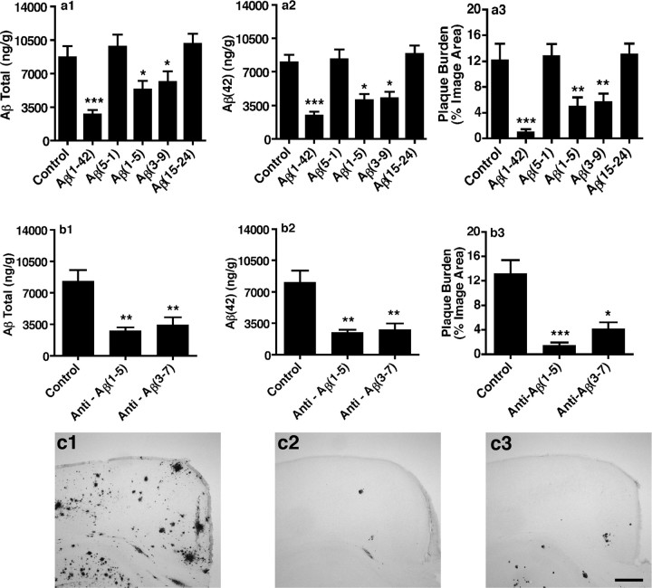
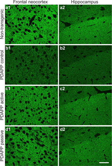
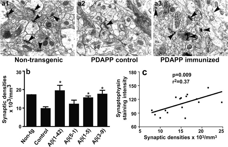
Alzheimer's disease neuropathology is characterized by key features that include the deposition of the amyloid beta peptide (Abeta) into plaques, the formation of neurofibrillary tangles, and the loss of neurons and synapses in specific brain regions. The loss of synapses, and particularly the associated presynaptic vesicle protein synaptophysin in the hippocampus and association cortices, has been widely reported to be one of the most robust correlates of Alzheimer's disease-associated cognitive decline. The beta-amyloid hypothesis supports the idea that Abeta is the cause of these pathologies. However, the hypothesis is still controversial, in part because the direct role of Abeta in synaptic degeneration awaits confirmation. In this study, we show that Abeta reduction by active or passive Abeta immunization protects against the progressive loss of synaptophysin in the hippocampal molecular layer and frontal neocortex of a transgenic mouse model of Alzheimer's disease. These results, substantiated by quantitative electron microscopic analysis of synaptic densities, strongly support a direct causative role of Abeta in the synaptic degeneration seen in Alzheimer's disease and strengthen the potential of Abeta immunotherapy as a treatment approach for this disease.


Figures

Figure 1.
Figure 1.
Active and passive Aβ immunization reduced Aβ levels and plaque burden in brains of the PDAPP mice. Brains of actively immunized (a1-a3), passively immunized (b1-b3), and control PDAPP mice were processed for measurements of total Aβ (a1, b1) and Aβ(42) (a2, b2) in the cortex by ELISA, or for measurement of plaque burden in the frontal neocortex (a3, b3, c1-c3) by quantitation of immunoperoxidase-stained brain sections (see Materials and Methods). Active immunization, either with full-length Aβ(1-42) or with N-terminal Aβ fragments, and passive immunization with N-terminal Aβ antibodies reduced levels of total Aβ and of Aβ(42) in the cortex of PDAPP mice. Similar results were obtained for the hippocampus(data not shown). The plaque burden in the frontal neocortex and hippocampus of a PDAPP mouse treated with either adjuvant alone (Control; c1), full-length Aβ(1-42) (Active; c2), or anti-Aβ(1-5) (Passive; c3) is shown. The results shown are means ± SEM. *p < 0.05, **p < 0.01, ***p < 0.001 (nonparametric Dunn's post hoc test; n = 16-25 mice per group). Scale bar, 0.3 mm.
Figure 2.
Figure 2.
Synaptic degeneration in PDAPP mice was age dependent. SYN-positive presynaptic terminal levels in the frontal neocortex (left) and hippocampal OML (Hippoc. Molec. Layer; right) of PDAPP mice and nontransgenic (Non-tg) littermate controls are shown. SYN levels were determined in immunofluorescent-labeled brain sections from 12- and 18-month-old PDAPP mice, as described in Materials and Methods. A significant SYN decrease was seen at 18 months, but not at 12 months, in both brain regions. The results shown are means ± SEM. ***p < 0.001 (Tukey's post hoc test; n = 10-17 mice per group).
Figure 3.
Figure 3.
Aβ immunization prevented synaptic degeneration in PDAPP mice. The images show SYN-positive presynaptic terminals in the frontal neocortex (left) and hippocampal OML (right) from an 18-month-old nontransgenic mouse (a1, a2), a control-treated PDAPP mouse (b1, b2), an actively immunized [Aβ(1-42)] PDAPP mouse (c1, c2), and a passively immunized (3D6) PDAPP mouse (d1, d2). Portions of two hippocampal subregions are visible in the right panels: the OML with a high synaptic density appears bright, and the stratum lacunosum with a lower synaptic density appears darker. Hippocampal SYN measurements were done in the OML. Note that SYN levels in immunized PDAPP mice are similar to those of nontransgenic mice. Scale bar, 50 μm.
Figure 4.
Figure 4.
Active and passive Aβ immunizations prevented SYN loss in the frontal neocortex and hippocampal OML (Hippoc. Molec. Layer) of PDAPP mice. For active immunization, 12-month-old PDAPP mice were treated with full-length Aβ [Aβ(1-42)] or with different Aβ fragments conjugates [Aβ(1-5),Aβ(3-9),Aβ(15-24)] for 6 months, as described in Materials and Methods. Control PDAPP mice received either vehicle only or a reverse peptide, Aβ(5-1). For passive immunization, 12-month-old PDAPP mice were treated for 6 months with either 3D6 [directed against Aβ(1-5)] or 12B4 [directed against Aβ(3-7)], dosed at 10 mg/kg for each weekly injection as described in Materials and Methods. Control PDAPP mice received injections of an irrelevant antibody or vehicle only. All antibodies were of the IgG2a isotype. SYN levels were analyzed in the frontal neocortex and hippocampal OML of 18-month-old PDAPP mice after completion of immunization. Significant improvements of SYN levels over controls were found after active immunization with Aβ(1-42), Aβ(1-5), and Aβ(3-9), but not Aβ(15-24) (a1, b1), and after passive immunization with either of two N-terminal Aβ antibodies (b1, b2). In these groups, SYN levels were not significantly different from that of nontransgenic mice or 12-month-old untreated PDAPP mice (see Figs. 2, 3). The results shown are means ± SEM. **p < 0.01, ***p < 0.001, by Dunnett's post hoc test (n = 16-25 mice per group).
Figure 5.
Figure 5.
SYN levels did not correlate with Aβ plaque burden. Correlation analyses were performed to determine whether loss of SYN correlated with plaque burden (both measured by quantitative immunohistochemistry; see Materials and Methods) in the frontal neocortex of PDAPP mice. To enhance the power of the analyses, all mice from the control groups of both active and passive immunization were grouped (a), and all mice from immunized groups that showed improvements of SYN levels over controls were grouped (b). There was no correlation between SYN levels and plaque burden in both cases. Similar results were obtained for the hippocampal OML (data not shown). This finding indicates that, in PDAPP mice, synapse loss is not reflected by measurements of total Aβ plaque burden. a, p = 0.48, r = 0.008, n = 58 mice; b, p = 0.42, r = -0.006, n = 95 mice. n.s., Not significant by Spearman rank correlation.
Figure 6.
Figure 6.
Ultrastructural quantification of neocortical synapses correlated with SYN levels in nontransgenic and in control-treated or immunized PDAPP mice. The images in a1-a3 show electron microscopic images of a neocortical area of a nontransgenic mouse (a1), a control (PBS)-treated PDAPP mouse (a2), and an actively immunized Aβ(1-5) PDAPP mouse (a3). The synaptic densities are marked by arrowheads. Note the diminished number of these densities in the PDAPP control compared with the nontransgenic mouse and the immunized PDAPP mouse. Magnification, 6000×. b, The bar graph shows the ultrastructural quantification (see Materials and Methods) of synaptic densities in nontransgenic (Non-tg; n = 2 mice), control (PBS)-treated PDAPP (n = 3 mice per group), and actively immunized [Aβ (1-42), Aβ (1-5), Aβ(3-9), or control Aβ(5-1)] PDAPP (n = 3 mice per group) mice. Note the significantly higher number of densities in immunized PDAPP mice compared with the two control groups (*p < 0.05; Dunnett's post hoc test). c, The line graph shows the correlation (Pearson's test) between the ultrastructural quantification of synaptic densities and the SYN levels obtained by quantitative immunohistochemistry (see Materials and Methods). Values from all animals examined by electron microscopy were pooled for the correlation analysis. Note the highly significant correlation between the two endpoints for synaptic integrity.

References

    1. Bard F, Cannon C, Barbour R, Burke RL, Games D, Grajeda H, Guido T, Hu K, Huang J, Johnson-Wood K, Khan K, Kholodenko D, Lee M, Lieberburg I, Motter R, Nguyen M, Soriano F, Vasquez N, Weiss K, Welch B, et al. (2000) Peripherally administered antibodies against amyloid beta-peptide enter the central nervous system and reduce pathology in a mouse model of Alzheimer disease. Nat Med 6: 916-919. - PubMed
    1. Bard F, Barbour R, Cannon C, Carretto R, Fox M, Games D, Guido T, Hoenow K, Hu K, Johnson-Wood K, Khan K, Kholodenko D, Lee C, Lee M, Motter R, Nguyen M, Reed A, Schenk D, Tang P, Vasquez N, et al. (2003) Epitope and isotype specificities of antibodies to beta-amyloid peptide for protection against Alzheimer's disease-like neuropathology. Proc Natl Acad Sci USA 100: 2023-2028. - PMC - PubMed
    1. Bussiere T, Bard F, Barbour R, Grajeda H, Guido T, Khan K, Schenk D, Games D, Seubert P, Buttini M (2004) Morphological characterization of thioflavin-S-positive amlyoid plaques in transgenic Alzheimer mice and effect of passive Abeta immunotherapy on their clearance. Am J Pathol 165: 987-995. - PMC - PubMed
    1. Butterfield DA, Bush AI (2004) Alzheimer's amyloid beta-peptide (1-42): involvement of methionine residue 35 in the oxidative stress and neurotoxicity properties of this peptide. Neurobiol Aging 25: 563-568. - PubMed
    1. Buttini M, Orth M, Bellosta S, Akeefe H, Pitas RE, Wyss-Coray T, Mucke L, Mahley RW (1999) Expression of human apolipoprotein E3 or E4 in the brains of Apoe-/-mice: isoform-specific effects on neurodegeneration. J Neurosci 19: 4867-4880. - PMC - PubMed

Publication types

MeSH terms