Comparative Study
. 2005 Oct 5;25(40):9176-84.
doi: 10.1523/JNEUROSCI.1632-05.2005.

Expression and modulation of an NADPH oxidase in mammalian astrocytes

Affiliations
Comparative Study

Expression and modulation of an NADPH oxidase in mammalian astrocytes

Andrey Y Abramov et al. J Neurosci. .

Abstract

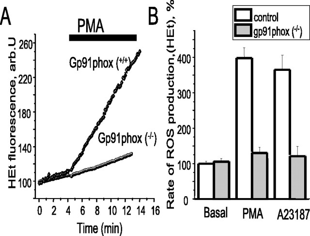
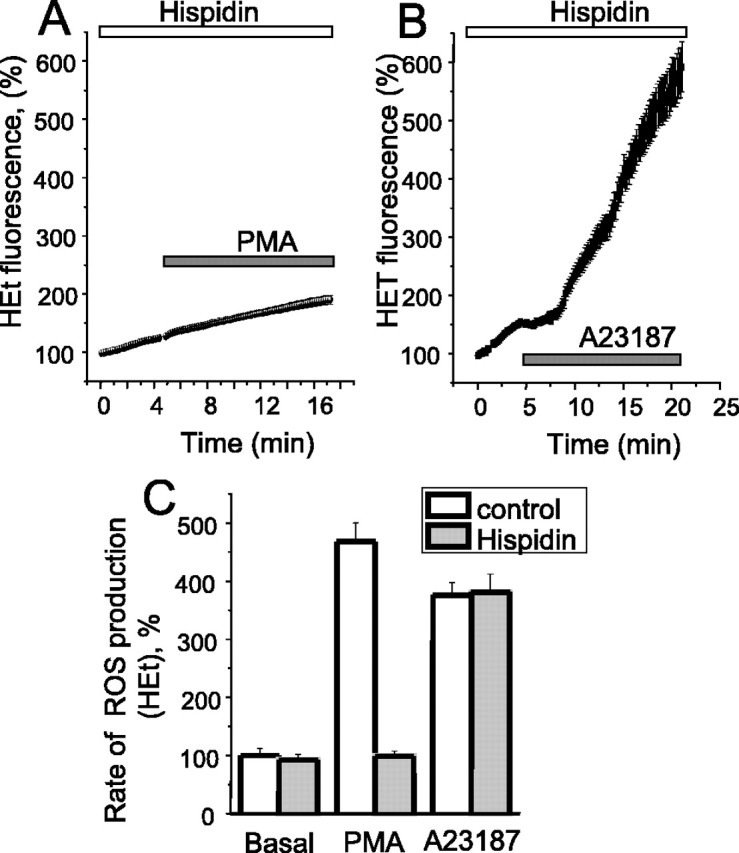
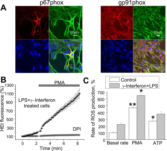
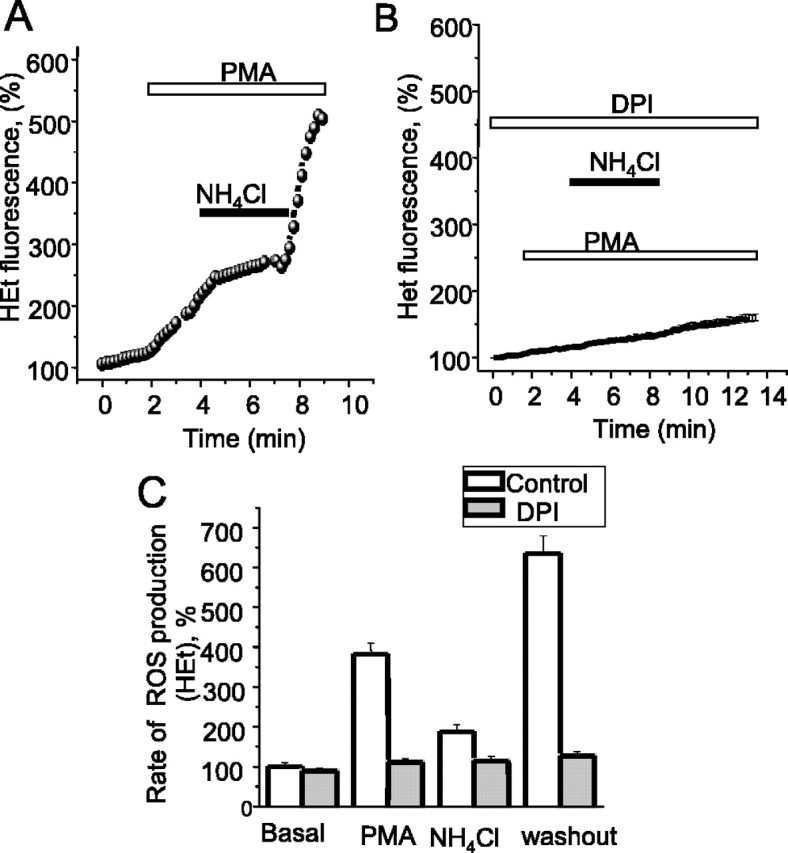
Amyloid beta peptides generate oxidative stress in hippocampal astrocytes through a mechanism sensitive to inhibitors of the NADPH oxidase [diphenylene iodonium (DPI) and apocynin]. Seeking evidence for the expression and function of the enzyme in primary hippocampal astrocytes, we confirmed the expression of the subunits of the phagocyte NADPH oxidase by Western blot analysis and by immunofluorescence and coexpression with the astrocyte-specific marker glial fibrillary acidic protein both in cultures and in vivo. Functional assays using lucigenin luminescence, dihydroethidine, or dicarboxyfluorescein fluorescence to measure the production of reactive oxygen species (ROS) demonstrated DPI and apocynin-sensitive ROS generation in response to the phorbol ester PMA and to raised [Ca2+]c after application of ionomycin or P2u receptor activation. Stimulation by PMA but not Ca2+ was inhibited by the protein kinase C (PKC) inhibitors staurosporine and hispidin. Responses were absent in transgenic mice lacking gp91phox. Expression of gp91phox and p67phox was increased in reactive astrocytes, which showed increased rates of both resting and stimulated ROS generation. NADPH oxidase activity was modulated by intracellular pH, suppressed by intracellular alkalinization, and enhanced by acidification. The protonophore carbonyl cyanide p-trifluoromethoxyphenylhydrazone suppressed basal ROS generation but markedly increased PMA-stimulated ROS generation. This was independent of mitochondrial ROS production, because it was unaffected by mitochondrial depolarization with rotenone and oligomycin. Thus, the NADPH oxidase is expressed in astrocytes and is functional, activated by PKC and intracellular calcium, modulated by pHi, and upregulated by astrocyte activation. The astrocytic NADPH oxidase is likely to play important roles in CNS physiology and pathology.


Figures

Figure 1.
Figure 1.
An NADPH oxidase is expressed in primary astrocytes. A, Western blots showing the expression of the subunits of the NADPH oxidase. i shows a Western blot of whole-cell extracts showing the expression of gp91phox and p67phox in astrocytes (a) using neutrophils (n) as a reference. PNS material from rat neutrophils and astrocytes was obtained and subjected to Western blot analysis as described in Materials and Methods. Six micrograms of neutrophil and 30 μg of astrocyte protein was applied and probed with antibodies to human gp91phox and p67phox as shown, because there is a high degree of homology between the proteins from human and rat. Arrowheads indicate the positions of the proteins. The figure is representative of three experiments. ii, Cytosol proteins probed for the cytosolic components of the NADPH oxidase, showing the expression of p67phox, p47phox, p40phox, and Rac as indicated. iii shows expression of the flavocytochrome subunits in the membrane fraction, namely gp91phox and p22phox as indicated. For a control and to ensure specificity, a control blot was made using antigp91phox antiserum, which was depleted for anti-gp91phox antibodies by adsorbing them out; this is not shown because the blot was completely blank. B, Immunofluorescence imaging using antibodies to gp91phox and p67phox counterstained with Cy5-labeled rabbit anti-human antibodies reveal the expression of both proteins in astrocyte cultures. Cells were counterstained with antibodies to GFAP (green) as a glial-specific marker and with DAPI to stain nuclei (blue), and images were acquired on a Zeiss (Welwyn Garden City, UK) 510 CLSM. These images are representative of five separate preparations, and staining was invariably seen clearly. The bottom panel shows a slightly higher gain image to show the distribution of the gp91phox. Appropriate controls showed no significant background signal under these conditions. Scale bars, 20 μm.
Figure 2.
Figure 2.
PMA stimulates ROS production by the NADPH oxidase in primary astrocyte cultures. Application of PMA induced an increase in ROS generation from astrocyte cultures measured using lucigenin luminescence (A, B), DCF (C), or HEt (D) fluorescence in response to 1 μg/ml PMA. The lucigenin response was reversed by the addition of superoxide dismutase to scavenge superoxide (A), demonstrating the specificity of the response, and was blocked by DPI (B). The gray traces in C and D show data from experiments in which cells were preincubated for 20 min with 0.5 μm DPI.
Figure 3.
Figure 3.
ROS production in astrocytes is activated by [Ca2+]c. A, The calcium ionophore ionomycin increased ROS generation, measured here with DCF fluorescence. B, Simultaneous measurements of [Ca2+]c (fura-2) and ROS production (HEt) in cortical astrocytes during application of 100 μm ATP. The mean values of the rate of ROS production measured with HEt are shown in C. In this case, signals measured from the rate of increase of HEt fluorescence were normalized to the rate at baseline taken as 100%. DPI (0.5 μm) or apocynin (1 mm) were used as inhibitors of NADPH oxidase (gray columns). D, Cells were bathed in a Ca2+-free saline containing 500 μm EGTA. The addition of thapsigargin (0.5 μm) released Ca2+ from the endoplasmic reticulum and caused small transient increase in ROS production while completely suppressing the response to subsequent application of ATP.
Figure 4.
Figure 4.
Astrocytes from gp91phox knock-out transgenic mice do not show increased ROS production in response to PMA or to Ca2+. Astrocyte cultures were prepared from a mouse exactly as described for the rat cells, using control and gp91 transgenic knock-out animals (a generous gift from D. Shah, King's College London, London, UK). Using HEt as described previously, the control cells showed a robust increase in ROS generation in response both to PMA (as illustrated in A; n = 58) and to an increase in [Ca2+] using A23187 (n = 39 cells; pooled data shown in B). In contrast, cells from the transgenic animals showed only minimal response to both stimuli (n = 60 for PMA and 58 for A23187). arb.U, Arbitrary units.
Figure 5.
Figure 5.
The PKCβ inhibitor hispidin inhibits PMA but not Ca2+-induced ROS production in cortical astrocytes. Cells were preincubated (20 min) with 20 μm hispidin. Hispidin inhibited the effect of 1 μg/ml PMA (A, C) but had no significant effect on the effect of the calcium ionophore 20 μm A23187 (B, C). In C, the mean values of the rates of ROS production measured with HEt are shown.
Figure 6.
Figure 6.
NADPH oxidase activity is upregulated in activated astrocyte (A) immunofluorescence images of gp91phox and p67phox (Cy5; red) counterstained with anti-GFAP antibodies (FITC; green), and DAPI (blue) shows that activated astrocytes have increased intensity of staining for GFAP and for both subunits of the oxidase. B, The increase in ROS production in response to PMA was significantly amplified after overnight treatment of astrocytes with LPS and γ-interferon (measured with HEt fluorescence). The histogram in C summarizes the rates of increase of HEt fluorescence in response to PMA with and without LPS and γ-interferon pretreatment.
Figure 7.
Figure 7.
Proton shunting with FCCP increases NADPH oxidase activity in cortical astrocytes. Application of 1 μm FCCP to control (A) cells caused a modest suppression of the basal rate of rise of HEt fluorescence. In contrast, application of FCCP to cortical astrocytes after PMA stimulation (B) caused a considerable increase in ROS generation. In the experiment presented in C, astrocytes were treated with 0.2 μg/ml oligomycin and 20 μm rotenone to cause complete mitochondrial depolarization before application of PMA. In this case, the additional application of FCCP still increased ROS generation, although the mitochondria were already depolarized.
Figure 8.
Figure 8.
Modulation of NADPH oxidase activity by intracellular pH. [pH]i was manipulated by proton loading using 5 mm NH4Cl. During NH4Cl exposure, the intracellular alkalosis was associated with a suppression of ROS generation, whereas the intracellular acidosis seen on NH4Cl washout enhanced ROS generation (A). All of these effects were abolished by DPI, confirming that these are effects of the changing pHi on oxidase activity and not because of pH sensitivity of the dyes (B). Pooled data are summarized in C.

References

    1. Abramov AY, Canevari L, Duchen MR (2003) Changes in [Ca2+]c and glutathione in astrocytes as the primary mechanism of amyloid neurotoxicity. J Neurosci 23: 5088-5095. - PMC - PubMed
    1. Abramov AY, Canevari L, Duchen MR (2004a) Amyloid β-peptides induce mitochondrial dysfunction and oxidative stress in astrocytes and death of neurons through activation of NADPH oxidase. J Neurosci 24: 565-575. - PMC - PubMed
    1. Abramov AY, Canevari L, Duchen MR (2004b) Calcium signals induced by amyloid β peptide and their consequences in neurons and astrocytes in culture. Biochim Biophys Acta 1742: 81-87. - PubMed
    1. Ahluwalia J, Tinker A, Clapp LH, Duchen MR, Abramov AY, Pope S, Nobles M, Segal AW (2004) The large-conductance Ca2+-activated K+ channel is essential for innate immunity. Nature 427: 853-858. - PMC - PubMed
    1. Banfi B, Molnar G, Maturana A, Steger K, Hegedus B, Demaurex N, Krause KH (2001) A Ca2+-activated NADPH oxidase in testis, spleen, and lymph nodes. J Biol Chem 276: 37594-38601. - PubMed

Publication types

MeSH terms

LinkOut - more resources