

Circadian regulation and function of voltage-dependent calcium channels in the suprachiasmatic nucleus
- PMID: 16207890
- PMCID: PMC6725766
- DOI: 10.1523/JNEUROSCI.2733-05.2005
Circadian regulation and function of voltage-dependent calcium channels in the suprachiasmatic nucleus
Abstract
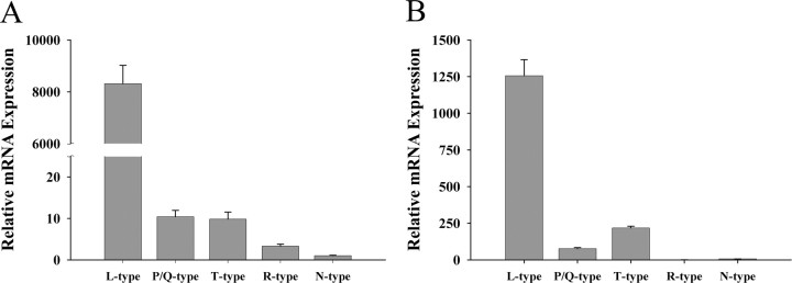
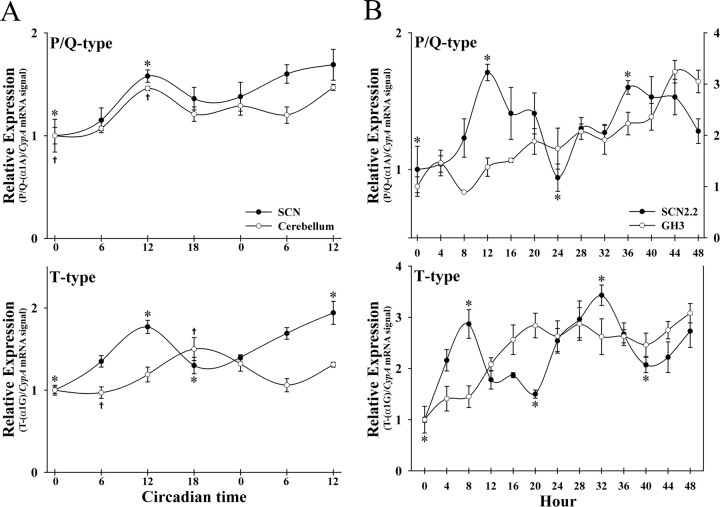
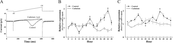
Individual neurons within the suprachiasmatic nuclei (SCNs) are capable of functioning as autonomous clocks and generating circadian rhythms in the expression of genes that form the molecular clockworks. Limited information is available on how these molecular oscillations in individual clock cells are coordinated to provide for the ensemble rhythmicity that is normally observed from the entire SCN. Because calcium influx via voltage-dependent calcium channels (VDCCs) has been implicated in the regulation of gene expression and synchronization of rhythmicity across the population of SCN clock cells, we first examined the rat SCN and an immortalized line of SCN cells (SCN2.2) for expression and circadian regulation of different VDCC alpha1 subunits. The rat SCN and SCN2.2 cells exhibited mRNA expression for all major types of VDCC alpha1 subunits. Relative levels of VDCC expression in the rat SCN and SCN2.2 cells were greatest for L-type channels, moderate for P/Q- and T-type channels, and minimal for R- and N-type channels. Interestingly, both rat SCN and SCN2.2 cells showed rhythmic expression of P/Q- and T-type channels. VDCC involvement in the regulation of molecular rhythmicity in SCN2.2 cells was then examined using the nonselective antagonist, cadmium. The oscillatory patterns of rPer2 and rBmal1 expression were abolished in cadmium-treated SCN2.2 cells without affecting cellular morphology and viability. These findings raise the possibility that the circadian regulation of VDCC activity may play an important role in maintaining rhythmic clock gene expression across an ensemble of SCN oscillators.
Figures
Similar articles
-
Oscillating on borrowed time: diffusible signals from immortalized suprachiasmatic nucleus cells regulate circadian rhythmicity in cultured fibroblasts.J Neurosci. 2001 Oct 15;21(20):7937-43. doi: 10.1523/JNEUROSCI.21-20-07937.2001. J Neurosci. 2001. PMID: 11588167 Free PMC article.
-
Daily rhythmicity of large-conductance Ca2+ -activated K+ currents in suprachiasmatic nucleus neurons.Brain Res. 2006 Feb 3;1071(1):54-62. doi: 10.1016/j.brainres.2005.11.078. Epub 2006 Jan 17. Brain Res. 2006. PMID: 16412396
-
Circadian profiling of the transcriptome in NIH/3T3 fibroblasts: comparison with rhythmic gene expression in SCN2.2 cells and the rat SCN.Physiol Genomics. 2007 May 11;29(3):280-9. doi: 10.1152/physiolgenomics.00199.2006. Epub 2007 Feb 6. Physiol Genomics. 2007. PMID: 17284666
-
Molecular analysis of mammalian circadian rhythms.Annu Rev Physiol. 2001;63:647-76. doi: 10.1146/annurev.physiol.63.1.647. Annu Rev Physiol. 2001. PMID: 11181971 Review.
-
Synchronization of the molecular clockwork by light- and food-related cues in mammals.Biol Chem. 2003 May;384(5):711-9. doi: 10.1515/BC.2003.079. Biol Chem. 2003. PMID: 12817467 Review.
Cited by
-
Calcium response to retinohypothalamic tract synaptic transmission in suprachiasmatic nucleus neurons.J Neurosci. 2007 Oct 24;27(43):11748-57. doi: 10.1523/JNEUROSCI.1840-07.2007. J Neurosci. 2007. PMID: 17959816 Free PMC article.
-
Stoichiometry of N-methyl-D-aspartate receptors within the suprachiasmatic nucleus.J Neurophysiol. 2010 Jun;103(6):3448-64. doi: 10.1152/jn.01069.2009. Epub 2010 Apr 21. J Neurophysiol. 2010. PMID: 20410362 Free PMC article.
-
Expanding clinical phenotype in CACNA1C related disorders: From neonatal onset severe epileptic encephalopathy to late-onset epilepsy.Am J Med Genet A. 2018 Dec;176(12):2733-2739. doi: 10.1002/ajmg.a.40657. Epub 2018 Dec 4. Am J Med Genet A. 2018. PMID: 30513141 Free PMC article.
-
Inhibition of p38 MAPK activity leads to cell type-specific effects on the molecular circadian clock and time-dependent reduction of glioma cell invasiveness.BMC Cancer. 2018 Jan 10;18(1):43. doi: 10.1186/s12885-017-3896-y. BMC Cancer. 2018. PMID: 29316898 Free PMC article.
-
Gut Microbiome-Brain Crosstalk in the Early Life of Chicken Reveals the Circadian Regulation of Key Metabolic and Immune Signaling Processes.Microorganisms. 2025 Mar 30;13(4):789. doi: 10.3390/microorganisms13040789. Microorganisms. 2025. PMID: 40284627 Free PMC article.
References
-
- Akasu T, Shoji S, Hasuo H (1993) Inward rectifier and low-threshold calcium currents contribute to the spontaneous firing mechanism in neurons of the rat suprachiasmatic nucleus. Pflügers Arch 425: 109-116. - PubMed
-
- Allen GC, Farnell Y, Bell-Pedersen D, Cassone VM, Earnest DJ (2004) Effects of altered Clock gene expression on the pacemaker properties of SCN2.2 cells and oscillatory properties of NIH/3T3 cells. Neuroscience 127: 989-999. - PubMed
-
- Baldelli P, Magnelli V, Carbone E (1999) Selective up-regulation of P- and R-type Ca2+ channels in rat embryonic motoneurons by BDNF. Eur J Neurosci 11: 1127-1133. - PubMed
-
- Catterall WA (2000) Structure and regulation of voltage-gated Ca2+ channels. Annu Rev Cell Dev Biol 16: 521-555. - PubMed
Publication types
MeSH terms
Substances
Grants and funding
LinkOut - more resources
Full Text Sources