

Rapid genotyping of hepatitis C virus by primer-specific extension analysis
- PMID: 16207978
- PMCID: PMC1248436
- DOI: 10.1128/JCM.43.10.5158-5163.2005
Rapid genotyping of hepatitis C virus by primer-specific extension analysis
Abstract
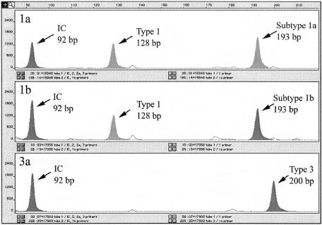
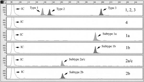
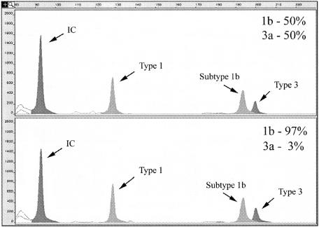
Quick and accurate genotyping of hepatitis C virus (HCV) is becoming increasingly important for clinical management of chronic infection and as an epidemiological marker. Furthermore, the incidence of HCV infection with mixed genotypes has clinical significance that is not addressed by most genotyping methods. We have developed a fluorescence-based genotyping assay called primer-specific extension analysis (PSEA) for the most prevalent HCV genotypes and have demonstrated the capacity of PSEA-HCV for detecting mixed-genotype HCV infections. PSEA-HCV detects genotype-specific sequence differences in the 5' untranslated region of HCV in products amplified by the COBAS AMPLICOR HCV Test, v2.0. Simulated mixed HCV infection of plasma with RNase-resistant RNA controls demonstrates that PSEA-HCV can detect as many as five genotypes in one specimen. Furthermore, in dual-genotype simulations, PSEA-HCV can unequivocally detect both genotypes, with one genotype representing only 3.1% of the mixture (313/10,000 IU in starting plasma). Compared to INNO-LiPA HCV II, both assays determined the same genotype for 191/199 (96%) patient specimens (175 subtype and 16 genotype-only identifications). Following the initial evaluation, PSEA-HCV was used routinely to genotype HCV from patient specimens submitted to our laboratory (n=312). Seventeen (5.4%) mixed infections were identified. The distribution of single-infection HCV genotypes in our population was 60.9% type 1 (n=190), 12.8% type 2 (n=40), 20.2% type 3 (n=63), 0.3% type 4 (n=1), and 0.3% other (n=1). In conclusion, PSEA-HCV provides an inexpensive, high-throughput screening tool for rapid genotyping of HCV while reliably identifying mixed HCV infections.
Figures
Similar articles
-
Evaluation of the invader assay for genotyping hepatitis C virus.J Clin Microbiol. 2006 Feb;44(2):318-23. doi: 10.1128/JCM.44.2.318-323.2006. J Clin Microbiol. 2006. PMID: 16455877 Free PMC article.
-
Determination of HCV genotype by direct sequence analysis of quantitative PCR products.J Med Virol. 2003 Feb;69(2):202-6. doi: 10.1002/jmv.10284. J Med Virol. 2003. PMID: 12683408
-
Detection of hepatitis C virus by a user-developed reverse transcriptase-PCR and use of amplification products for subsequent genotyping.J Clin Virol. 2004 Oct;31(2):148-52. doi: 10.1016/j.jcv.2004.02.010. J Clin Virol. 2004. PMID: 15364272
-
Profile of Roche's cobas® HCV tests.Expert Rev Mol Diagn. 2017 Apr;17(4):311-319. doi: 10.1080/14737159.2017.1293526. Epub 2017 Feb 22. Expert Rev Mol Diagn. 2017. PMID: 28277143 Review.
-
Profile of the VERSANT HCV genotype 2.0 assay.Expert Rev Mol Diagn. 2018 Dec;18(12):995-1004. doi: 10.1080/14737159.2018.1541740. Epub 2018 Nov 2. Expert Rev Mol Diagn. 2018. PMID: 30372355 Review.
Cited by
-
Characterization of hepatitis C virus genotypes by direct sequencing of HCV 5'UTR region of isolates from Saudi Arabia.PLoS One. 2014 Aug 6;9(8):e103160. doi: 10.1371/journal.pone.0103160. eCollection 2014. PLoS One. 2014. PMID: 25099694 Free PMC article. Clinical Trial.
-
A public health response to a newly diagnosed case of hepatitis C associated with lapse in Infection Prevention and Control practices in a dental setting in Ontario, Canada.Can Commun Dis Rep. 2021 Jul 8;47(7-8):347-352. doi: 10.14745/ccdr.v47i78a08. eCollection 2021 Jul 8. Can Commun Dis Rep. 2021. PMID: 34421388 Free PMC article.
-
CIHR Canadian HIV Trials Network Coinfection and Concurrent Diseases Core Research Group: 2016 Updated Canadian HIV/Hepatitis C Adult Guidelines for Management and Treatment.Can J Infect Dis Med Microbiol. 2016;2016:4385643. doi: 10.1155/2016/4385643. Epub 2016 Jul 4. Can J Infect Dis Med Microbiol. 2016. PMID: 27471521 Free PMC article.
-
An update on the management of chronic hepatitis C: 2015 Consensus guidelines from the Canadian Association for the Study of the Liver.Can J Gastroenterol Hepatol. 2015 Jan-Feb;29(1):19-34. doi: 10.1155/2015/692408. Epub 2015 Jan 13. Can J Gastroenterol Hepatol. 2015. PMID: 25585348 Free PMC article.
-
Burden of disease and cost of chronic hepatitis C infection in Canada.Can J Gastroenterol Hepatol. 2014 May;28(5):243-50. doi: 10.1155/2014/317623. Can J Gastroenterol Hepatol. 2014. PMID: 24839620 Free PMC article. Review.
References
-
- Bukh, J., R. H. Miller, and R. H. Purcell. 1995. Genetic heterogeneity of hepatitis C virus: quasispecies and genotypes. Semin. Liver Dis. 15:41-63. - PubMed
-
- Germer, J. J., D. W. Majewski, M. Rosser, A. Thompson, P. S. Mitchell, T. F. Smith, S. Elagin, and J. D. C. Yao. 2003. Evaluation of the TRUGENE HCV 5′NC genotyping kit with the new GeneLibrarian module 3.1.2 for genotyping of hepatitis C virus from clinical specimens. J. Clin. Microbiol. 41:4855-4857. - PMC - PubMed
Publication types
MeSH terms
Substances
LinkOut - more resources
Full Text Sources
Other Literature Sources
