. 2005 Oct 6;437(7060):902-5.
doi: 10.1038/nature04147.

STIM1 is a Ca2+ sensor that activates CRAC channels and migrates from the Ca2+ store to the plasma membrane

Affiliations

STIM1 is a Ca2+ sensor that activates CRAC channels and migrates from the Ca2+ store to the plasma membrane

Shenyuan L Zhang et al. Nature. .

Abstract

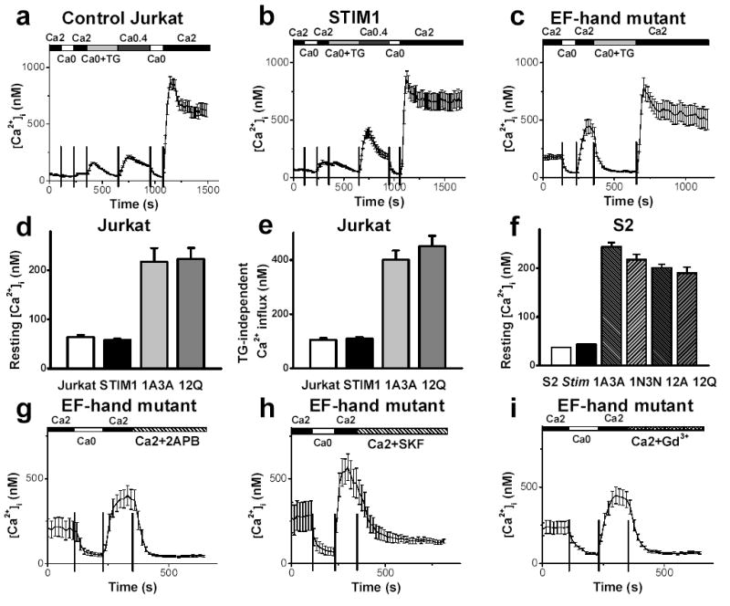
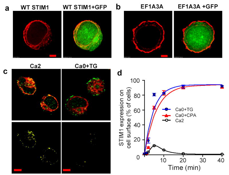
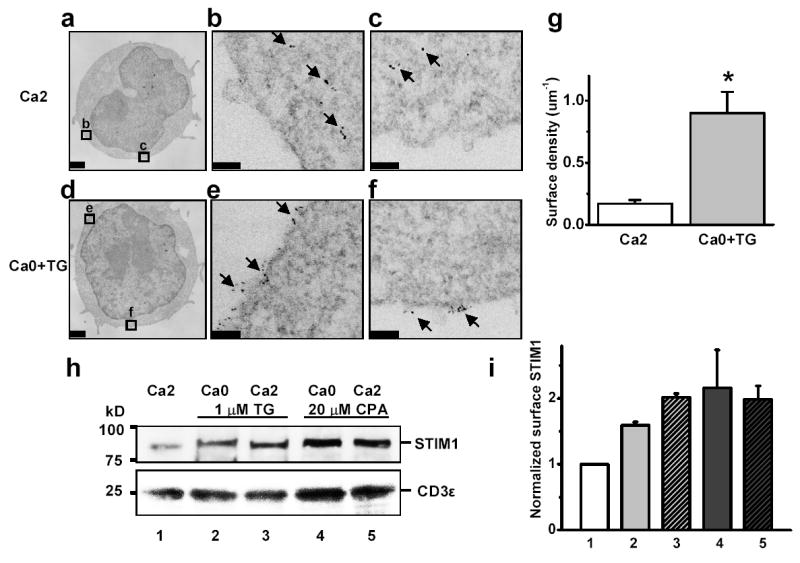
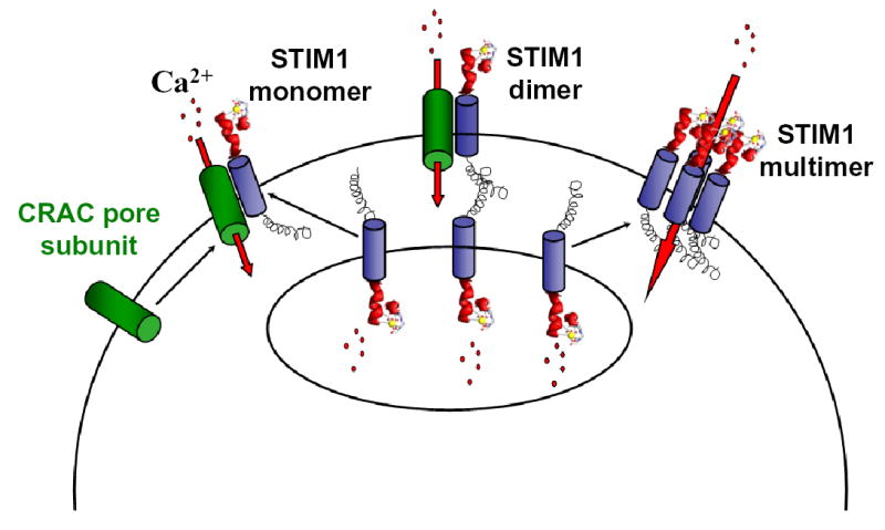
As the sole Ca2+ entry mechanism in a variety of non-excitable cells, store-operated calcium (SOC) influx is important in Ca2+ signalling and many other cellular processes. A calcium-release-activated calcium (CRAC) channel in T lymphocytes is the best-characterized SOC influx channel and is essential to the immune response, sustained activity of CRAC channels being required for gene expression and proliferation. The molecular identity and the gating mechanism of SOC and CRAC channels have remained elusive. Previously we identified Stim and the mammalian homologue STIM1 as essential components of CRAC channel activation in Drosophila S2 cells and human T lymphocytes. Here we show that the expression of EF-hand mutants of Stim or STIM1 activates CRAC channels constitutively without changing Ca2+ store content. By immunofluorescence, EM localization and surface biotinylation we show that STIM1 migrates from endoplasmic-reticulum-like sites to the plasma membrane upon depletion of the Ca2+ store. We propose that STIM1 functions as the missing link between Ca2+ store depletion and SOC influx, serving as a Ca2+ sensor that translocates upon store depletion to the plasma membrane to activate CRAC channels.


Figures

Figure 1
Figure 1. Constitutive activation of CRAC channels by expression of EF hand mutant of Stim or STIM1
a–c, Average [Ca2+]i responses in blank-transfected Jurkat cells (n=59), Jurkat cells overexpressing WT STIM1 (n=29), and Jurkat cells overexpressing STIM1 EF12Q (n=18), respectively. Cells were bathed in solutions as indicated. d–f, Resting [Ca2+]i in Jurkat cells (from left to right: n=114, n=171, n=61, and n=65); TG-independent Ca2+ influx in Jurkat cells (n=114, n=136, n=61, and n=42); and resting [Ca2+]i in S2 cells (from left to right: n=392, n=568, n=425, n=149, n=316, and n=102). g–i, Effects of CRAC channel blockers 2-APB (50 μM), SKF96365 (20 μM) and Gd3+ (1 μM) on TG-independent Ca2+ influx in Jurkat cells. EF hand mutants: 12Q (g, n=16), 12Q (h, n=8), and 1A3A (i, n=28). Error bars: SEM.
Figure 2
Figure 2. Mutations in EF hand motif or store depletion induce STIM1 translocation to plasma membrane
a,b, STIM1 (red) immunofluorescence staining of Jurkat cells transfected with WT STIM1 (a) or EF1A3A STIM1 (b), in 2 mM Ca2+ Ringer solution. GFP (green) was co-transfected to define the cytoplasmic region. Scale bar is 2 μm. c, STIM1 (green) and SERCA2 (red) immunofluorescence staining of Jurkat cells in 2 mM Ca2+ Ringer solution or 1 μM TG in zero-Ca2+ solution for 10 min. Note the separation of STIM1 at the cell surface (top right). Bottom panels: colocalization images (yellow) that depict pixels that contained both STIM1 and ER fluorescence. Note reduced olocalization caused by STIM1 translocation in TG-treated cells. Scale bar is 5 μm. d, Time course of STIM1 translocation triggered by 1 μM TG or 10 μM CPA, represented by the percentage of cells with strong surface fluorescence. An average of 315 cells was counted for each time point. Time constants: 4.7 min (TG), 6.1 min (CPA). Error bars: SEM.
Figure 3
Figure 3. Subcellular distribution of STIM1 before and after store depletion: immuno-electron microscopy and surface biotinylation
a, Quantum dot-labeled STIM1 in control Jurkat cells bathed in 2 mM Ca2+ Ringer solution. b,c, Enlargements of corresponding boxed areas in a. d, STIM1 in Jurkat cells pretreated with 1 μM TG in zero-Ca2+ solution for 15 min. e,f, Enlarged regions showing clustered STIM1 distribution on plasma membrane. Scale bar is 1 μm for a and d; 0.1 μm for b,c,e,f. g, STIM1 surface density, pooled data from control cells (n = 14) and store-depleted cells (n = 13); asterisk denotes P value < 0.0005 compared to controls. h, Jurkat cells were treated for 30 min as indicated. Biotinylated proteins were detected by antibodies to STIM1 (top) and CD3ɛ (bottom). i, Quantification of biotinylation experiments: 1.6–2.2 fold increase upon store depletion. Error bars: SEM.
Figure 4
Figure 4. Models of STIM1 Function
Upon store depletion, STIM1 located in the ER unbinds Ca2+ and translocates to the plasma membrane to activate CRAC channel subunits that are already in the plasma membrane (left), form junctions between the Ca2+ store and the plasma membrane (middle), or assemble to form functional CRAC channels (right).

References

    1. Putney JW, Jr, Broad LM, Braun FJ, Lievremont JP, Bird GS. Mechanisms of capacitative calcium entry. J Cell Sci. 2001;114:2223–9. - PubMed
    1. Putney JW., Jr Store-operated calcium channels: how do we measure them, and why do we care? Sci STKE. 2004;2004:pe37. - PubMed
    1. Lewis RS. Store-operated calcium channels. Adv Second Messenger Phosphoprotein Res. 1999;33:279–307. - PubMed
    1. Parekh AB, Putney JW., Jr Store-operated calcium channels. Physiol Rev. 2005;85:757–810. - PubMed
    1. Lewis RS, Cahalan MD. Mitogen-induced oscillations of cytosolic Ca2+ and transmembrane Ca2+ current in human leukemic T cells. Cell Regul. 1989;1:99–112. - PMC - PubMed

Publication types

MeSH terms