Review
. 2005 Oct;2(5):719-29.
doi: 10.1586/14789450.2.5.719.

Histone structure and nucleosome stability

Affiliations
Review

Histone structure and nucleosome stability

Leonardo Mariño-Ramírez et al. Expert Rev Proteomics. 2005 Oct.

Abstract

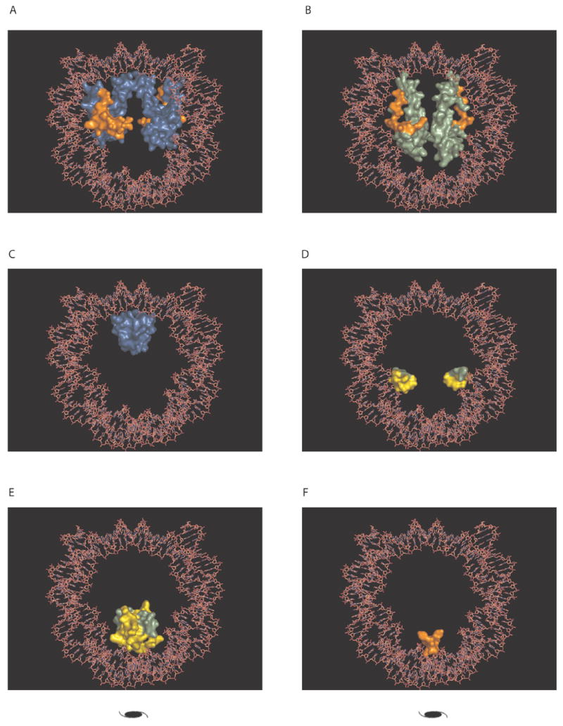
Histone proteins play essential structural and functional roles in the transition between active and inactive chromatin states. Although histones have a high degree of conservation due to constraints to maintain the overall structure of the nucleosomal octameric core, variants have evolved to assume diverse roles in gene regulation and epigenetic silencing. Histone variants, post-translational modifications and interactions with chromatin remodeling complexes influence DNA replication, transcription, repair and recombination. The authors review recent findings on the structure of chromatin that confirm previous interparticle interactions observed in crystal structures.


Figures

Figure 1
Figure 1. Post-translational modifications in the nucleosome core particle near the protein-DNA interface
(A) The nucleosome core particle structure (Protein Data Bank accession code 1KX5) with the histone modifications that could be involved in increasing nucleosome mobility. The colours represent different histones H3 (blue), H4 (light green), H2A (orange) and H2B (yellow); the histone modifications are shown as spheres. (B) The histone fold domain of H3and H4 interacting in the characteristic handshake motif. The figure was prepared with PyMCL [102].
Figure 2
Figure 2. Electrostatic surface potential of the nucleosome core particle (Protein Data Bank accession code 1KX5)
The electrostatic potential ranges from +20 (blue) to −20 (red) kTe−1. (A) Side view of the nucleosome core particle. Note the distribution of positive charges (blue) around the DNA interface and the core histone tails. (B) Top view of the nucleosome core particle. This surface has a number of negative charges (red) indicated by a green arrow in the exposed protein surface of the histone octamer. The electrostatic potential mapped to the molecular surface was calculated using Graphical Representation and Analysis of Structural Properties (GRASP) [69], and the figures were prepared with the Swiss-PDB Viewer [70].
Figure 3
Figure 3. Schematic representation of the regions of the core histone sequences in pairs of interacting chains
Each core histone is represented by a black line drawn to sequence length scale, the location of interacting residues is indicated by colored boxes depending on the interacting mode (see TABLE 2 for details; each mode in TABLE 2 is identified). The box encompasses all residues in the region of contact. Specific residues are identified in TABLE 2. aa: Amino acids.
Figure 4
Figure 4. Conserved regions of histories H4 andH2A, which are the contacts for internucleosomal interactions
Sequence logo representation of multiple sequence alignments of histoneH4 and H2A. The multiple sequence alignments were obtained from the Histone Database [101]; secondary structure elements are shown as lines (loops) and boxes (helices). The amino adds that make contacts in the crystal structures are marked above the sequence logos. (A) Histone H4 alignment around the basic region that mediates internucleosomal interactions. Lysine 16 and 20 can be subject to post-translational modifications. (B) HistoneH2Aalignment around the addle region that mediates internucleosomal interactions. The figure was prepared with WebLogo [71].
Figure 5
Figure 5. Conserved binding modes in a representative nucleosome core particle structure
TABLE 2 describes the specific interface residues involved in the conserved interaction. Core histones are represented as molecular surf aces and are colored the same as for FIGURE 1: histone H3 (blue), H4 (light green), H2A (orange) and H2B (yellow). The crystallographic dyad is indicated by a dyad symbol. (A) Conserved binding mode 3, interaction between H3 and H2A (B) Conserved binding mode 4, interaction between H4 and H2A (C) Conserved binding mode 5, interaction between H3 and H3. (D) Conserved binding mode 6, interaction between H4 and H2B (E) Conserved binding mode 7, interaction between H4 and H2B (F) Conserved binding mode 8, interaction between H2A and H2A The figure was prepared with PyMOL[102].

References

    1. Luger K, Mader AW, Richmond RK, Sargent DF, Richmond TJ. Crystal structure of the nucleosome core particle at 2.8 Å resolution. Nature. 1997;389(6648):251–260. - PubMed
    2. •• Contains high-resolution structures of the nucleosome that are key to understanding gene regulation and chromatin remodeling.

    1. Davey CA, Sargent DF, Luger K, Maeder AW, Richmond TJ. Solvent mediated interactions in the structure of the nucleosome core particle at 1.9 Å resolution. J Mol Biol. 2002 ;319(5):1097–1113. - PubMed
    2. •• Contains high-resolution structures of the nucleosome that are key to understanding gene regulation and chromatin remodeling.

    1. Dorigo B, Schalch T, Kulangara A, et al. Nucleosome arrays reveal the two-start organization of the chromatin fiber. Science. 2004;306(5701):1571–1573. - PubMed
    2. • Provides evidence for a two-start helical organization of the 30-nm chromatin fiber and has helped resolve a controversy over how individual nucleosomes are packed in the chromatin fiber.

    1. van Holde KE. Chromatin. Springer-Verlag; London, UK: 1988. Chapter 7: Highter order structure; pp. 317–354.
    1. Francis NJ, Kingston RE, Woodcock CL. Chromatin compaction by a polycomb group protein complex. Science. 2004;306(5701):1574–1577. - PubMed

Websites

    1. Marino-Ramirez L, Hsu B, Baxevanis AD, Landsman D NHGRI/NCBI. http://research.nhgri.nih.gov/histones (Viewed September 2005)
    1. Delano WL. The PyMOL Molecular Graphics System. DeLano Scientific; San Carlos, CA, USA: 2002. http://www.pymol.org (Viewed September 2005)