

Cerebral processing of painful oesophageal stimulation: a study based on independent component analysis of the EEG
- PMID: 16210400
- PMCID: PMC1856147
- DOI: 10.1136/gut.2005.068460
Cerebral processing of painful oesophageal stimulation: a study based on independent component analysis of the EEG
Abstract
Background and aims: Independent component analysis (ICA) of the electroencephalogram (EEG) overcomes many of the classical problems in EEG analysis. We used ICA to determine the brain responses to painful stimulation of the oesophagus.
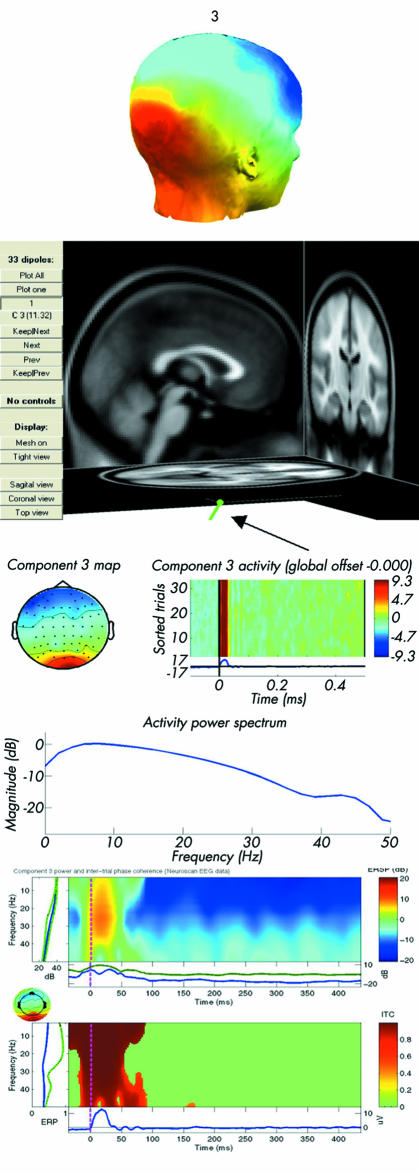
Methods: Twelve subjects with a median age of 41 years were included. With a nasal endoscope, two series of 35 electrical stimuli at the pain threshold were given to the distal oesophagus and the EEG was subjected to ICA. The sessions were separated by 30 minutes. For each component head models, event related images, spectral perturbation, coherence analysis, and dipoles were extracted. The most valid components were found according to time/frequency information and reliability in both experiments.
Results: Reliable components with the most valid dipoles were found in the thalamus, insula, cingulate gyrus, and sensory cortex. Time locked activities were consistent with upstream activation of these areas, and cross coherence analysis of the sources demonstrated dynamic links in the beta(14-25 Hz) and gamma(25-50 Hz) bands between the suggested networks of neurones. The thalamic components were time and phase locked intermittently, starting around 50 ms. In the cingulate gyrus, the posterior areas were always firstly activated, followed by the middle and anterior regions. Components with dipoles in the sensory cortex were localised in several regions of the somatosensory area.
Conclusions: The method gives new information relating to the localisation and dynamics between neuronal networks in the brain to pain evoked from the human oesophagus, and should be used to increase our understanding of clinical pain.
Conflict of interest statement
Conflict of interest: None declared.


Comment in
-
Independent component analysis of the EEG: is this the way forward for understanding abnormalities of brain-gut signalling?Gut. 2006 May;55(5):597-600. doi: 10.1136/gut.2005.081703. Gut. 2006. PMID: 16609130 Free PMC article.
Similar articles
-
Differences in perception and brain activation following stimulation by large versus small area cutaneous surface electrodes.Eur J Pain. 2012 Jul;16(6):827-37. doi: 10.1002/j.1532-2149.2011.00063.x. Epub 2011 Dec 19. Eur J Pain. 2012. PMID: 22337577 Clinical Trial.
-
Adaptation in human somatosensory cortex as a model of sensory memory construction: a study using high-density EEG.Brain Struct Funct. 2016 Jan;221(1):421-31. doi: 10.1007/s00429-014-0915-5. Epub 2014 Oct 29. Brain Struct Funct. 2016. PMID: 25352155
-
Real-time imaging of human cortical activity evoked by painful esophageal stimulation.Gastroenterology. 2005 Mar;128(3):610-9. doi: 10.1053/j.gastro.2004.12.033. Gastroenterology. 2005. PMID: 15765397
-
Measuring our natural painkiller.Trends Neurosci. 2002 Feb;25(2):67-8; discussion 69. doi: 10.1016/s0166-2236(02)02025-8. Trends Neurosci. 2002. PMID: 11814550 Review.
-
Electrophysiological Signature of Pain.Adv Exp Med Biol. 2018;1099:167-177. doi: 10.1007/978-981-13-1756-9_15. Adv Exp Med Biol. 2018. PMID: 30306524 Review.
Cited by
-
Altered brain microstructure assessed by diffusion tensor imaging in patients with diabetes and gastrointestinal symptoms.Diabetes Care. 2013 Mar;36(3):662-8. doi: 10.2337/dc12-1131. Epub 2012 Nov 8. Diabetes Care. 2013. PMID: 23139372 Free PMC article.
-
Electroencephalography and analgesics.Br J Clin Pharmacol. 2014 Jan;77(1):72-95. doi: 10.1111/bcp.12137. Br J Clin Pharmacol. 2014. PMID: 23593934 Free PMC article. Review.
-
Independent component analysis of the EEG: is this the way forward for understanding abnormalities of brain-gut signalling?Gut. 2006 May;55(5):597-600. doi: 10.1136/gut.2005.081703. Gut. 2006. PMID: 16609130 Free PMC article.
-
The pain system in oesophageal disorders: mechanisms, clinical characteristics, and treatment.Gastroenterol Res Pract. 2011;2011:910420. doi: 10.1155/2011/910420. Epub 2011 Aug 2. Gastroenterol Res Pract. 2011. PMID: 21826137 Free PMC article.
-
Is the pain in chronic pancreatitis of neuropathic origin? Support from EEG studies during experimental pain.World J Gastroenterol. 2008 Jul 7;14(25):4020-7. doi: 10.3748/wjg.14.4020. World J Gastroenterol. 2008. PMID: 18609686 Free PMC article.
References
Publication types
MeSH terms
LinkOut - more resources
Full Text Sources
Other Literature Sources
Medical
Miscellaneous