. 2005 Oct;12(10):1177-83.
doi: 10.1128/CDLI.12.10.1177-1183.2005.

Serologic cross-reactivity between Anaplasma marginale and Anaplasma phagocytophilum

Affiliations

Serologic cross-reactivity between Anaplasma marginale and Anaplasma phagocytophilum

U M Dreher et al. Clin Diagn Lab Immunol. 2005 Oct.

Abstract

In the context of a serosurvey conducted on the Anaplasma marginale prevalence in Swiss cattle, we suspected that a serological cross-reactivity between A. marginale and A. phagocytophilum might exist. In the present study we demonstrate that cattle, sheep and horses experimentally infected with A. phagocytophilum not only develop antibodies to A. phagocytophilum (detected by immunofluorescent-antibody assay) but also to A. marginale (detected by a competitive enzyme-linked immunosorbent assay). Conversely, calves experimentally infected with A. marginale also developed antibodies to A. phagocytophilum using the same serological tests. The identity of 63% determined in silico within a 209-amino-acid sequence of major surface protein 5 of an isolate of A. marginale and one of A. phagocytophilum supported the observed immunological cross-reactivity. These observations have important consequences for the serotesting of both, A. marginale and A. phagocytophilum infection of several animal species. In view of these new findings, tests that have been considered specific for either infection must be interpreted carefully.


Figures
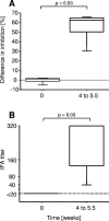
FIG. 1.
FIG. 1.
Antibody detection in cELISA (A) and IFA (B) of five calves experimentally infected with A. marginale. Samples were taken from each animal prior to (week 0) and after infection (weeks 4 to 5.5). Antibody test results in samples collected after infection were significantly higher than in preinfection serum samples (P = 0.03) which is indicated.
FIG. 2.
FIG. 2.
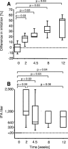
Sera from six cows, prior to and after experimental infection with A. phagocytophilum. Antibodies measured in cELISA (A) to detect A. marginale and A. centrale antibodies and in IFA (B) designed to quantitate A. phagocytophilum antibodies in the same serum samples. The mean inhibitory value at week zero was set as baseline in panel A. Significant differences as indicated.
FIG. 3.
FIG. 3.
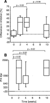
Antibody values measured as inhibitory values in cELISA (A) and as titers in IFA (B) of five sheep experimentally infected with A. phagocytophilum. The mean inhibition before infection at week zero was set as baseline in panel A. Significant differences as indicated.
FIG. 4.
FIG. 4.
Antibody quantification by cELISA (A) and IFA (B) in sera from six horses experimentally infected with A. phagocytophilum. Samples were taken prior to (day 0) and after infection (day 12). Results of samples collected at day 0 were significantly different from those of samples collected at day 12, in both, the IFA and in the cELISA (both P = 0.02). The inhibitory values of cELISA shown in panel A were set to reach 0% inhibition in the noninfected horses.
FIG. 5.
FIG. 5.
Western blot analysis. Antigen produced in tick cells (Ixodes scapularis embryonic cells, IDE8 cells, infected with A. marginale Virginia isolate). Ladders: 25, 20, 15, and 10 kDa. Lanes: 1, negative control serum included in the cELISA test; 2, positive control serum included in the cELISA test; 3, monoclonal antibody against MSP5; 4, serum of horse two, taken before infection; 5, serum of horse two, taken 12 days after infection with A. phagocytophilum. The band of 18 kDa (right arrow) detected by the positive control serum (2) and monoclonal antibody (3) is also detected in the horse after infection with A. phagocytophilum (5). Many bands are detected in both negative and positive control sera, presumably representing antibodies against tick cell antigens. The monoclonal antibody specific for A. marginale MSP5 recognized not only MSP5 but also a band of 15 kDa which was recognized by antibodies present in several sera collected from noninfected animals. No explanation was found for this MAb to bind to a band, which most likely is of tick cell origin.
FIG. 6.
FIG. 6.
Alignment of Anaplasma sp. MSP5 protein sequences. Conserved amino acids are shaded in gray and amino acid positions conserved in three or four of five sequences are shaded in black.

References

    1. Asanovich, K. M., J. S. Bakken, J. E. Madigan, M. Aguero-Rosenfeld, G. P. Wormser, and J. S. Dumler. 1997. Antigenic diversity of granulocytic Ehrlichia isolates from humans in Wisconsin and New York and a horse in California. J. Infect. Dis. 176:1029-1034. - PubMed
    1. de la Fuente, J., E. J. Golsteyn Thomas, R. A. van den Bussche, R. G. Hamilton, E. E. Tanaka, S. E. Druhan, and K. M. Kocan. 2003. Characterization of Anaplasma marginale isolated from North American bison. Appl. Environ. Microbiol. 69:5001-5005. - PMC - PubMed
    1. de la Fuente, J., R. F. Massung, S. J. Wong, F. K. Chu, H. Lutz, M. Meli, F. D. von Loewenich, A. Grzeszczuk, A. Torina, S. Caracappa, A. J. Mangold, V. Naranjo, S. Stuen, and K. M. Kocan. 2005. Sequence analysis of the msp4 gene of Anaplasma phagocytophilum strains. J. Clin. Microbiol. 43:1309-1317. - PMC - PubMed
    1. Dreher, U. M., R. Hofmann-Lehmann, M. L. Meli, G. Regula, A. Y. Cagienard, K. D. Stark, M. Doherr, F. Filli, M. Hässig, U. Braun, K. M. Kocan, and H. Lutz. 2005. Seroprevalence of anaplasmosis among cattle in Switzerland in 1998 and 2003: no evidence of an emerging disease. Vet. Microbiol. 107:71-79. - PubMed
    1. Dumler, J. S., A. F. Barbet, C. P. Bekker, G. A. Dasch, G. H. Palmer, S. C. Ray, Y. Rikihisa, and F. R. Rurangirwa. 2001. Reorganization of genera in the families Rickettsiaceae and Anaplasmataceae in the order Rickettsiales: unification of some species of Ehrlichia with Anaplasma, Cowdria with Ehrlichia and Ehrlichia with Neorickettsia, descriptions of six new species combinations and designation of Ehrlichia equi and ‘HGE agent’ as subjective synonyms of Ehrlichia phagocytophila. Int. J. Syst. Evol. Microbiol. 51:2145-2165. - PubMed

Publication types

Substances

LinkOut - more resources