

Bcr is a negative regulator of the Wnt signalling pathway
- PMID: 16211085
- PMCID: PMC1371031
- DOI: 10.1038/sj.embor.7400536
Bcr is a negative regulator of the Wnt signalling pathway
Abstract
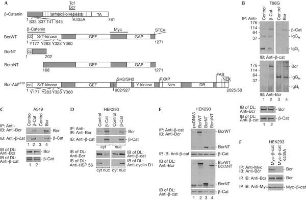
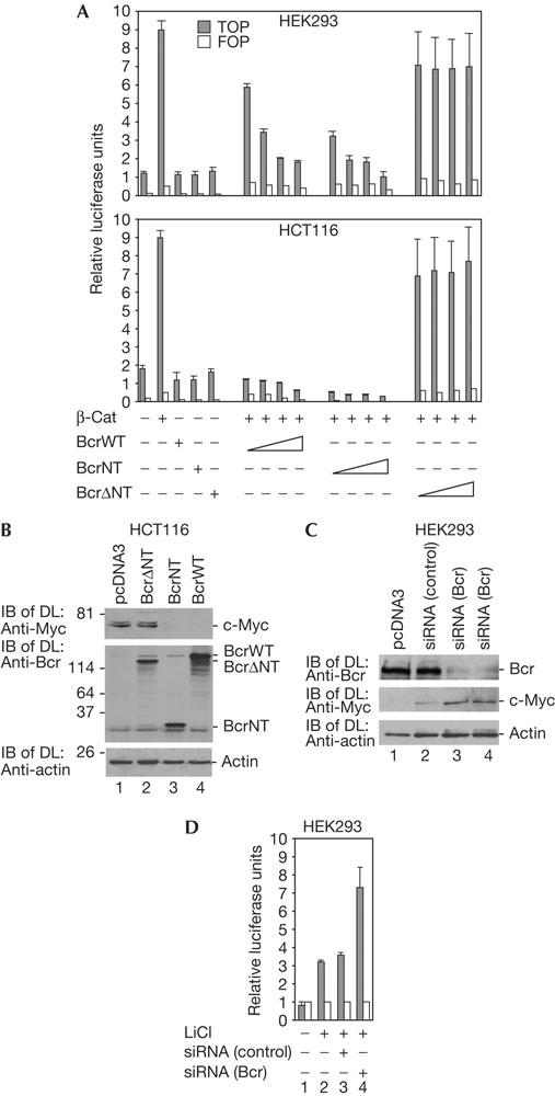
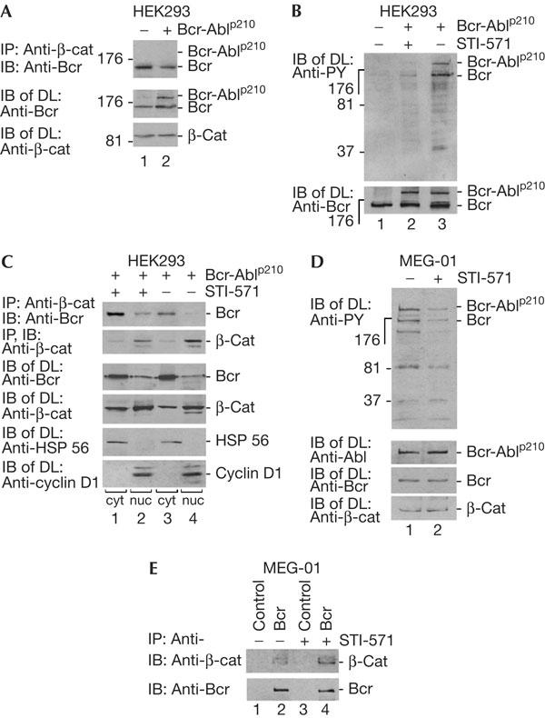
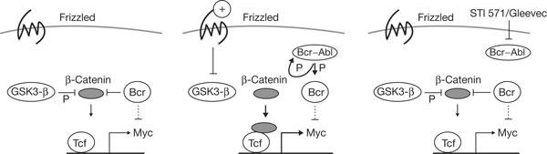
The Wnt signalling pathway can activate transcription of genes such as c-myc through beta-catenin. Here, we describe the protein breakpoint cluster region, Bcr, as a negative regulator of this pathway. Bcr can form a complex with beta-catenin and negatively regulate expression of c-Myc. Knockdown of Bcr by short interfering RNA relieves the block and activates expression of c-Myc. Expression of Bcr in the human colon carcinoma cell line HCT116, which has a high level of endogenous beta-catenin, leads to reduced c-Myc expression. The negative effect is exerted by the amino terminus of Bcr, which does not harbour the kinase domain. Bcr-Abl, the oncogene protein expressed in chronic myelogenous leukaemia (CML), does not bind to beta-catenin. It phosphorylates Bcr in the first exon sequence on tyrosines, which abrogates the binding of Bcr to beta-catenin. The inhibitor of the Bcr-Abl tyrosine kinase, STI-571 or Gleevec, a drug against CML, reverses this effect. Our data contribute to the understanding of Bcr as a tumour suppressor in the Wnt signalling pathway, as well as in CML.
Figures
Similar articles
-
Bcr-Abl stabilizes beta-catenin in chronic myeloid leukemia through its tyrosine phosphorylation.EMBO J. 2007 Mar 7;26(5):1456-66. doi: 10.1038/sj.emboj.7601485. Epub 2007 Feb 22. EMBO J. 2007. PMID: 17318191 Free PMC article.
-
Downregulation of γ-catenin inhibits CML cell growth and potentiates the response of CML cells to imatinib through β-catenin inhibition.Int J Mol Med. 2013 Feb;31(2):453-8. doi: 10.3892/ijmm.2012.1207. Epub 2012 Dec 6. Int J Mol Med. 2013. PMID: 23233089
-
Janus kinase 2: a critical target in chronic myelogenous leukemia.Cancer Res. 2006 Jul 1;66(13):6468-72. doi: 10.1158/0008-5472.CAN-06-0025. Cancer Res. 2006. PMID: 16818614
-
Chronic myelogenous leukaemia--new therapeutic principles.J Intern Med. 2001 Jul;250(1):3-9. doi: 10.1046/j.1365-2796.2001.00823.x. J Intern Med. 2001. PMID: 11454136 Review.
-
Mutated tyrosine kinases as therapeutic targets in myeloid leukemias.Adv Exp Med Biol. 2003;532:121-40. doi: 10.1007/978-1-4615-0081-0_11. Adv Exp Med Biol. 2003. PMID: 12908554 Review.
Cited by
-
Genetic/molecular alterations of meningiomas and the signaling pathways targeted.Oncotarget. 2015 May 10;6(13):10671-88. doi: 10.18632/oncotarget.3870. Oncotarget. 2015. PMID: 25965831 Free PMC article. Review.
-
Ubiquitin-mediated interaction of p210 BCR/ABL with β-catenin supports disease progression in a murine model for chronic myelogenous leukemia.Blood. 2013 Sep 19;122(12):2114-24. doi: 10.1182/blood-2013-01-481184. Epub 2013 Aug 15. Blood. 2013. PMID: 23950177 Free PMC article.
-
Inhibition of DDR1-BCR signalling by nilotinib as a new therapeutic strategy for metastatic colorectal cancer.EMBO Mol Med. 2018 Apr;10(4):e7918. doi: 10.15252/emmm.201707918. EMBO Mol Med. 2018. PMID: 29438985 Free PMC article.
-
DDR1 contributes to kidney inflammation and fibrosis by promoting the phosphorylation of BCR and STAT3.JCI Insight. 2022 Feb 8;7(3):e150887. doi: 10.1172/jci.insight.150887. JCI Insight. 2022. PMID: 34941574 Free PMC article.
-
An RNA-seq protocol to identify mRNA expression changes in mouse diaphyseal bone: applications in mice with bone property altering Lrp5 mutations.J Bone Miner Res. 2013 Oct;28(10):2081-93. doi: 10.1002/jbmr.1946. J Bone Miner Res. 2013. PMID: 23553928 Free PMC article.
References
-
- Arlinghaus RB (2002) Bcr: a negative regulator of the Bcr–Abl oncoprotein in leukemia. Oncogene 21: 8560–8567 - PubMed
-
- Daley GQ, Van Etten RA, Baltimore D (1990) Induction of chronic myelogenous leukemia in mice by the P210bcr/abl gene of the Philadelphia chromosome. Science 247: 824–830 - PubMed
-
- Giles RH, van Es JH, Clevers H (2003) Caught up in a Wnt storm: Wnt signaling in cancer. Biochim Biophys Acta 1653: 1–24 - PubMed
Publication types
MeSH terms
Substances
LinkOut - more resources
Full Text Sources
Miscellaneous
