. 2005 Nov;115(11):3276-84.
doi: 10.1172/JCI24685. Epub 2005 Oct 6.

The role of 2 FOXP3 isoforms in the generation of human CD4+ Tregs

Affiliations

The role of 2 FOXP3 isoforms in the generation of human CD4+ Tregs

Sarah E Allan et al. J Clin Invest. 2005 Nov.

Abstract

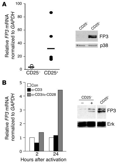
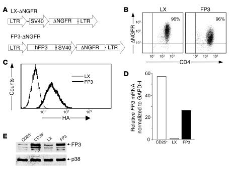
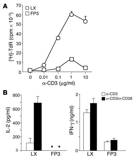
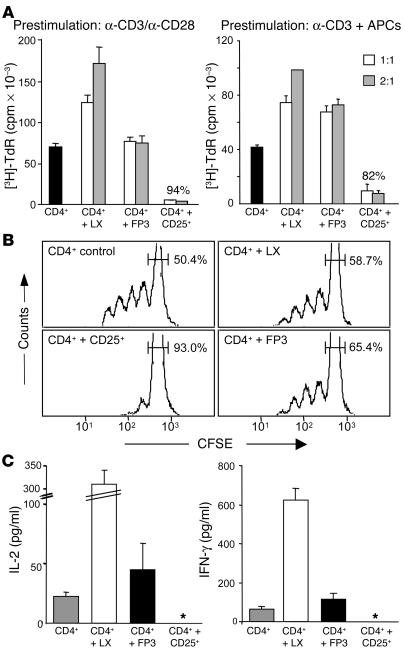
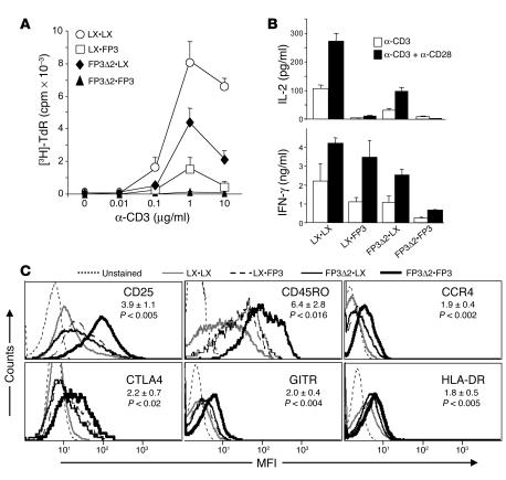
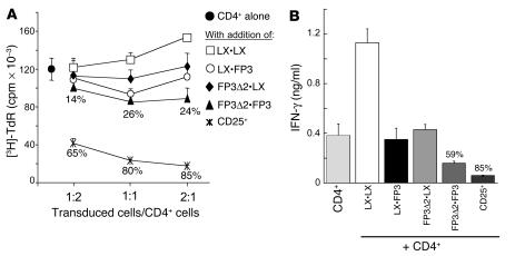
Little is known about the molecules that control the development and function of CD4+ CD25+ Tregs. Recently, it was shown that the transcription factor FOXP3 is necessary and sufficient for the generation of CD4+ CD25+ Tregs in mice. We investigated the capacity of FOXP3 to drive the generation of suppressive CD4+ CD25+ Tregs in humans. Surprisingly, although ectopic expression of FOXP3 in human CD4+ T cells resulted in induction of hyporesponsiveness and suppression of IL-2 production, it did not lead to acquisition of significant suppressor activity in vitro. Similarly, ectopic expression of FOXP3delta2, an isoform found in human CD4+ CD25+ Tregs that lacks exon 2, also failed to induce the development of suppressor T cells. Moreover, when FOXP3 and FOXP3delta2 were simultaneously overexpressed, although the expression of several Treg-associated cell surface markers was significantly increased, only a modest suppressive activity was induced. These data indicate that in humans, overexpression of FOXP3 alone or together with FOXP3delta2 is not an effective method to generate potent suppressor T cells in vitro and suggest that factors in addition to FOXP3 are required during the process of activation and/or differentiation for the development of bona fide Tregs.


Figures

Figure 1
Figure 1
Endogenous expression of FOXP3 (FP3) in human CD4+ T cells. CD4+ T cells were purified and separated into CD25 and CD25+ cells. (A) Expression of FOXP3 was determined by quantitative RT-PCR, with each point representing an individual donor, and Western blotting. (B) Highly purified CD4+CD25 T cells were activated with immobilized anti-CD3 (1 μg/ml) and/or anti-CD28 (1 μg/ml) for the indicated times and assayed for mRNA expression. Levels of FOXP3 protein were determined after 3 days in cells left unstimulated (–) or stimulated (+) with anti-CD3 and anti-CD28. Protein levels were compared with those in ex vivo CD4+CD25+ Tregs. Results are representative of 5 experiments. Con, control.
Figure 2
Figure 2
Ectopic expression of FOXP3 in human CD4+CD25CD45RA+ T cells. (A) Control- (LX-) or FOXP3- encoding retroviruses expressing ΔNGFR as a marker gene were used for transduction of CD4+CD25CD45RA+ T cells. (B) Following purification, transduced T cells were analyzed by flow cytometry. (C) In some cases, T cells were transduced with HA-tagged FOXP3, and coexpression of ΔNGFR and FOXP3 was monitored by analysis of HA expression in ΔNGFR+ T cells. (D) Quantitative RT-PCR and (E) Western blotting were performed to determine expression levels of FOXP3. Ex vivo CD4+CD25 and CD4+CD25+ T cells were included to compare relative levels of expression. Results are representative of at least 8 tests with T cells derived from 3 different donors.
Figure 3
Figure 3
Proliferative capacity and cytokine production profile of FOXP3-transduced T cells. (A) Transduced T cells were tested for their ability to proliferate in response to increasing concentrations of immobilized anti-CD3 mAbs, based on incorporation of tritiated thymidine ([3H]TdR). (B) T cells were stimulated with anti-CD3 mAbs (1 μg/ml), with or without anti-CD28 mAbs (1 μg/ml), for 24 hours (IL-2) and 48 hours (IFN-γ). The asterisk indicates less than 9 pg/ml. Culture supernatants were analyzed by ELISA. Data are representative of 5 independent experiments for A and 3 for B.
Figure 4
Figure 4
Ectopic expression of FOXP3 is not sufficient for suppressive function. (A) Autologous CD4+ T cells were stimulated with soluble anti-CD3 mAbs (1 μg/ml) and APCs, in the presence or absence of control- (LX-) or FOXP3-transduced T cells, which were generated in the absence or presence of APCs, at a 1:1 or 2:1 (transduced cells/target cells) ratio. Ex vivo–isolated CD4+CD25+ Tregs were added as a positive control. (B) In parallel experiments, proliferation of CFSE-labeled autologous CD4+ T cells, alone or in the presence of a 1:1 ratio of control- (LX-) or FOXP3-transduced T cells, or ex vivo CD4+CD25+ (CD25+) Tregs was analyzed by flow cytometry after 96 hours. Numbers represent the percentage of undivided cells in the cultures. (C) Culture supernatants were collected and analyzed by cytometric bead assay to determine amounts of IL-2 (after 36 hours) or IFN-γ (after 72 hours). Results are representative of 4 independent experiments for A and 3 for B and C. The asterisks indicate less than 9 pg/ml.
Figure 5
Figure 5
Two isoforms of FOXP3 in human CD4+CD25+ Tregs with repressor activity. (A) RNA was extracted from purified CD4+CD25+ cells of normal donors and analyzed by RT-PCR using primers specific for a region of FOXP3 spanning exons 1 and 3. (B) Jurkat T cells were cotransfected with an hIL-2–luciferase reporter, β-galactosidase, and either control- (LX-), FOXP3-, or FOXP3Δ2- (FP3Δ2-) encoding plasmids. Following stimulation with PMA and Ca2+ ionophore, luciferase activity was determined and normalized to amounts of β-galactosidase activity. The graph depicts the percent activity of the reporter in comparison to the LX-transfected control. Data represent the average of 5 independent experiments.
Figure 6
Figure 6
Retroviral transduction of CD4+CD25CD45RA+ T cells with FOXP3Δ2 in the absence or presence of FOXP3. (A) Control- (LX-) or FOXP3Δ2-encoding retroviral vectors expressing GFP as a marker gene were used in combination with ΔNGFR-expressing retroviral vectors (see Figure 2A) for double transduction of CD4+CD25CD45RA+ T cells. Following FACS sorting and expansion, transduced T cells were analyzed by flow cytometry (B), quantitative RT-PCR (C), and Western blotting (D). Results are representative of T cells derived from 3 different donors.
Figure 7
Figure 7
Proliferative capacity, cytokine production, and cell-surface phenotype of cells expressing FOXP3Δ2 in the absence or presence of FOXP3. (A) Transduced T cells were tested for their ability to proliferate in response to increasing concentrations of immobilized anti-CD3 mAbs. (B) T cells were stimulated with anti-CD3 mAbs (1 μg/ml), with or without anti-CD28 mAbs (1 μg/ml), for 24 hours (IL-2) and 48 hours (IFN-γ). Culture supernatants were analyzed by ELISA. (C) Expression levels of the indicated proteins was determined in the resting phase. Numbers indicate the average fold increase in MFI in the FP3Δ2•FP3 T cells compared with the LX•LX controls. Data are representative of a minimum of 6 independent experiments with T cells derived from 2 different donors.
Figure 8
Figure 8
Coexpression of FOXP3 and FOXP3Δ2 is not sufficient for acquisition of suppressor activity at levels equivalent to CD4+CD25+ Tregs. (A) Autologous CD4+ T cells were stimulated with soluble anti-CD3 mAbs (1 μg/ml) and APCs, in the presence or absence of double-transduced T cells at increasing transduced cell/target cell ratios. Ex vivo–isolated CD4+CD25+ Tregs were added as a positive control. (B) Culture supernatants were collected and analyzed by cytometric bead assay to determine amounts of IFN-γ after 72 hours. Numbers represent the percent suppression compared with CD4+ T cells alone. For A, data are representative of 3 experiments where suppression at a 1:1 ratio was observed (out of a total of 7). B is representative of 4 independent experiments.

Similar articles

Cited by

References

    1. Levings MK, Bacchetta R, Schulz U, Roncarolo MG. The role of IL-10 and TGF-beta in the differentiation and effector function of T regulatory cells. Int. Arch. Allergy Immunol. 2002;129:263–276. - PubMed
    1. Sakaguchi S. Naturally arising CD4+ regulatory T cells for immunologic self-tolerance and negative control of immune responses. Annu. Rev. Immunol. 2004;22:531–562. - PubMed
    1. Shevach EM, McHugh RS, Piccirillo CA, Thornton AM. Control of T-cell activation by CD4+ CD25+ suppressor T cells. Immunol. Rev. 2001;182:58–67. - PubMed
    1. Levings MK, Sangregorio R, Roncarolo MG. Human CD25+CD4+ T regulatory cells suppress naive and memory T-cell proliferation and can be expanded in vitro without loss of function. J. Exp. Med. 2001;193:1295–1302. - PMC - PubMed
    1. Dieckmann D, Plottner H, Berchtold S, Berger T, Schuler S. Ex vivo isolation and characterization of CD4+CD25+ T cells with regulatory properties from human blood. J. Exp. Med. 2001;193:1303–1310. - PMC - PubMed

Publication types

MeSH terms