. 2005 Nov;115(11):3205-16.
doi: 10.1172/JCI24179. Epub 2005 Oct 6.

Germinal center exclusion of autoreactive B cells is defective in human systemic lupus erythematosus

Affiliations

Germinal center exclusion of autoreactive B cells is defective in human systemic lupus erythematosus

Amedeo Cappione 3rd et al. J Clin Invest. 2005 Nov.

Abstract

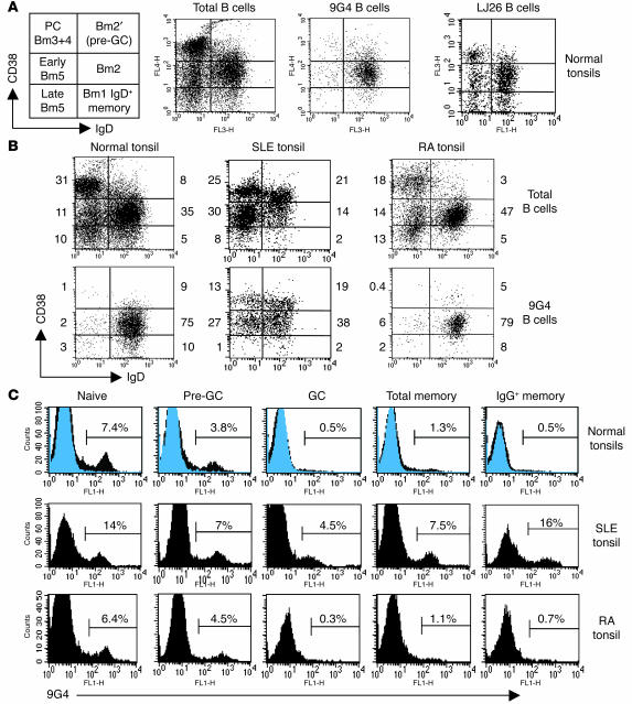
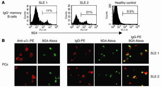
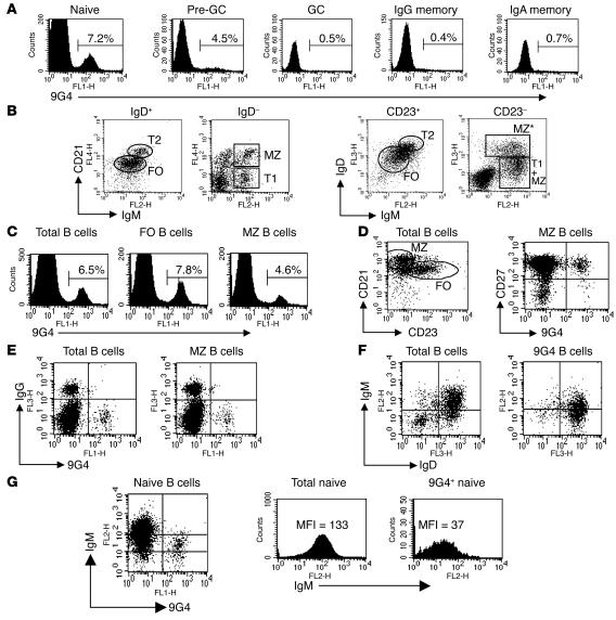
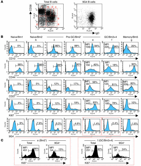
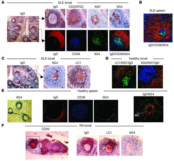
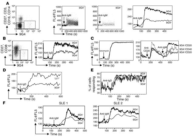
Breach of B cell tolerance is central to the pathogenesis of systemic lupus erythematosus (SLE). However, how B cell tolerance is subverted in human SLE is poorly understood due to difficulties in identifying relevant autoreactive B cells and in obtaining lymphoid tissue. We have circumvented these limitations by using tonsil biopsies to study autoreactive B cells (9G4 B cells), whose regulation is abnormal in SLE. Here we show that 9G4 B cells are physiologically excluded during the early stages of the GC reaction before acquiring a centroblast phenotype. Furthermore, we provide evidence to indicate that an anergic response to B cell receptor stimulation may be responsible for such behavior. In contrast, in SLE, 9G4 B cells progressed unimpeded through this checkpoint, successfully participated in GC reactions, and expanded within the post-GC IgG memory and plasma cell compartments. The faulty regulation of 9G4 B cells was not shared by RA patients. To our knowledge, this work represents the first comparative analysis of the fate of a specific autoreactive human B cell population. The results identify a defective tolerance checkpoint that appears to be specific for human SLE.


Figures

Figure 1
Figure 1
9G4 cells actively participate in GC reactions in SLE. (A) IgD/CD38 expression identifies the populations shown in the diagram on the left: IgD+CD38 (Bm1 and IgD+ memory); IgD+CD38+ (Bm2); IgD+CD38++ (Bm2′ pre-GC cells); IgDCD38++ (Bm3 and Bm4 or centroblasts and centrocytes); IgDCD38+ (early Bm5 memory); IgDCD38 (late Bm5 memory); and CD38bright plasma cells (PC). Dot plots show the typical distribution of total, 9G4, and control VH3 (LJ26) B cells in normal tonsils. In every tonsil, 9G4 B cells were significantly underrepresented in the GC and memory compartments. (B) Tonsillar B cells from normal controls and SLE and RA patients analyzed as described above. Total tonsillar B cells demonstrate naive lymphopenia and expansion of pre-GC cells described in studies of SLE peripheral blood (68). In SLE, 9G4 B cells are not blocked at the pre-GC stage. Instead, they are greatly expanded in the GC and memory compartments. Yet these abnormalities are not found in tonsillar B cells from RA patients where 9G4 cells are normally distributed. The percentage of each subpopulation is shown on the margins of the dot plots. (C) Histograms depict the frequency of 9G4 cells in normal, SLE, and RA tonsils. SLE patients show a large increase in the frequency of 9G4 GC cells and a 10- to 25-fold increase in IgG 9G4 memory cells compared with healthy controls and RA patients. Blue histograms represent the staining obtained with the corresponding isotype control antibodies (data shown for normal controls), while 9G4 histograms are depicted in black.
Figure 2
Figure 2
9G4 B cells are expanded in the post-GC compartments. (A) FACS analysis of PBL CD19+ B cells from active SLE patients stained with CD27, IgG, and 9G4 antibodies. A large increase in IgG 9G4 memory cells was demonstrated in SLE as compared with the very low levels typical of healthy subjects. Two representative SLE examples are shown. (B) For these same patients, CD138+ PCs were analyzed for cytoplasmic expression of the 9G4 idiotype and light chains or IgG. A large fraction of both total and IgG plasma cells in SLE were 9G4+.
Figure 3
Figure 3
Analysis of 9G4 cells in healthy spleens. (A) CD19+ spleen B cells were analyzed with IgD, CD38, CD27, and 9G4 antibodies as described above (n = 7 spleens). 9G4 B cells are very scarce within the GC and post-GC compartments (IgG and IgA memory). Representative results are shown as histograms. (B) Staining for IgM, IgD, CD21, and CD23 expression identified transitional (T1 and T2), follicular (FO), and MZ populations with a distribution similar to mouse B cells (12, 69). We identified an additional fraction composed of significant numbers of IgD+ cells, which represents a distinct subset of IgD+ MZ B cells (MZ*) (19). (C) Total spleen B cells were fractionated into MZ and follicular subsets as described above and further analyzed for the frequency of 9G4 B cells. The frequency of spleen follicular 9G4 cells was similar to the tonsil, and a lower but significant frequency was observed in the MZ fraction. (D) The majority of total and 9G4 MZ B cells express CD27. (E) The dearth of IgG and IgA 9G4 B cells was consistently documented in the spleen whether using total B cells or fractionated MZ B cells. (F) Dot plot analysis of total and 9G4 spleen B cells demonstrated that the vast majority of 9G4 B cells express an IgM+IgD+ phenotype. (G) Within the naive compartment, 9G4 B cells express significantly lower levels of surface IgM. MFI, mean fluorescence intensity.
Figure 4
Figure 4
9G4 cells are normally censored at the GC founder stage. (A) The left dot plot is representative of normal tonsils, demonstrating a prominent GC founder population (fraction e). As shown in the dot plot on the right, even in these tonsils, 9G4 B cells fail to progress past the pre-GC compartment and are scarce among GC founders. (B) Tonsils were analyzed for the expression of developmental markers CD10, CD44, and CD27 on conventional Bm1–Bm5 subsets (fractions a, b, f, and g). Pre-GC/Bm2′ cells were further divided into 3 fractions (c–e), with e containing the putative GC founders. As shown in the corresponding histograms, CD10 (a GC marker) was progressively acquired in fractions c–f, while CD44 (a marker downregulated in GC) was progressively lost. CD27 also experienced progressive upregulation in fractions c–f. Strikingly, the highest expression of the nuclear proliferation protein Ki67 was observed in fraction e. These results are consistent with fraction e representing GC founders undergoing the initial phases of clonal expansion. Importantly, the majority of 9G4 B cells was lost during pre-GC progression, greatly underrepresented among GC founders, and failed to expand within the GC, where their frequency continued to decline. (C) The scarce 9G4 B cells present within the GC founder and GC compartments were further analyzed for intracellular Ki67 expression. Consistent with their inability to form productive GC reactions, and in contrast to total B cells within these fractions, 9G4 B cells expressed very low levels of Ki67.
Figure 5
Figure 5
9G4 GCs are present in SLE patients at high frequency. (A) SLE tonsil biopsies stained with anti-IgD (left). Mature GCs formed by expansions of 9G4 cells were frequently identified in SLE. The upper row of the enlarged images depicts a typical mature 9G4+ proliferative GC with a well-formed FDC network (CD23/FDC) and positive Ki67 staining. The lower row shows similar findings by immunofluorescence: follicular mantle (IgD-PE, red), GC (CD38–7-aminomethylcoumarin, blue), and 9G4 (Alexa 488, green). (B) Representative example of a 9G4+ GC in an SLE spleen shown by 3-color immunofluorescence. (C) SLE GC showing the expansion of both 9G4 and LC1 B cells. (D) Healthy tonsils stained by immunofluorescence: IgD (red), Ki67 (blue), and either 9G4 or LC1 (both green). In contrast to SLE, healthy tonsils lack 9G4+ GC. Yet proliferating LC1 GCs were readily demonstrated. (E) A representative field from healthy spleens demonstrating the absence of 9G4 staining in the GCs is shown on the left. These results were routinely corroborated by immunofluorescence (middle panels). The photograph on the right illustrates the absence of 9G4 B cells from the GC and their accumulation within the FO and the MZ. (F) Enzymatic staining of serial sections obtained from tonsil biopsies of patients with RA failed to demonstrate 9G4+ GCs. Instead, as in healthy subjects, 9G4 B cells were restricted to the follicular mantle. In contrast, LC1+ GCs were readily identified in these patients.
Figure 6
Figure 6
9G4 B cells display attenuated calcium responses. (A) Calcium responses in healthy PBL B cells after anti-IgM stimulation. B cells were purified by negative selection and stained with CD27-, CD3-, CD14-, and CD16-PE antibodies (to gate out non-B cells and memory B cells) and 9G4 at the time of calcium measurements (gating shown on the left). The responses of individual cells are depicted in the middle dot plots, and the median responses in the graphs at right. (B) Calcium responses in tonsillar naive B cells after anti-IgD stimulation. Cells were stained with CD38 mAb and CD27 mAb to gate out memory and GC B cells, as well as 9G4 at the time of calcium measurements. The attenuated response of 9G4+ cells is not due to IgM downmodulation, since similar results were seen with anti-IgD. (C) 9G4 B cells normally mobilize calcium in response to ionomycin and CD20 cross-linking. Yet anti-CD20 failed to restore full signaling upon subsequent BCR stimulation. Of note, CD40 costimulation by overnight incubation with CD40 ligand–expressing fibroblasts (dotted line) increased the response through both CD20 and the BCR, yet it failed to fully restore BCR signaling to the same level seen for 9G4 cells. (D) 9G4 naive B cells have attenuated signaling compared with another positively selected naive B cell population. 9G4 tonsillar B cells were purified by magnetic selection using 9G4-FITC. Control VH4 naive B cells were purified using LC1. Calcium responses in the naive gated population were examined as described above. (E) The percentage of cells responding to BCR stimulation was similar in 9G4+ cells as compared with naive control B cells, indicating that the differences shown in D were due to lower average response per cell. (F) Calcium signaling through the BCR is similarly attenuated in PBL naive 9G4 B cells from SLE patients.

Similar articles

Cited by

References

    1. Hochberg MC. Updating the American College of Rheumatology revised criteria for the classification of systemic lupus erythematosus [letter] Arthritis Rheum. 1997;40:1725. - PubMed
    1. Winfield JB, Winchester RJ, Kunkel HG. Association of cold-reactive antilymphocyte antibodies with lymphopenia in systemic lupus erythematosus. Arthritis Rheum. 1975;18:587–594. - PubMed
    1. Cappione AJ, Pugh-Bernard AE, Anolik JH, Sanz I. Lupus IgG VH4.34 antibodies bind to a 220-kDa glycoform of CD45/B220 on the surface of human B lymphocytes. J. Immunol. 2004;172:4298–4307. - PubMed
    1. Chan OT, Madaio MP, Shlomchik MJ. The central and multiple roles of B cells in lupus pathogenesis. Immunol. Rev. 1999;169:107–121. - PubMed
    1. Pugh-Bernard AE, et al. Regulation of inherently autoreactive VH4-34 B cells in the maintenance of human B cell tolerance. J. Clin. Invest. 2001;108:1061–1070. - PMC - PubMed

Publication types

MeSH terms