. 2005 Nov;115(11):3157-65.
doi: 10.1172/JCI23475. Epub 2005 Oct 6.

Pemphigus foliaceus IgG causes dissociation of desmoglein 1-containing junctions without blocking desmoglein 1 transinteraction

Affiliations

Pemphigus foliaceus IgG causes dissociation of desmoglein 1-containing junctions without blocking desmoglein 1 transinteraction

Jens Waschke et al. J Clin Invest. 2005 Nov.

Abstract

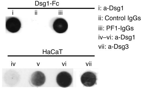
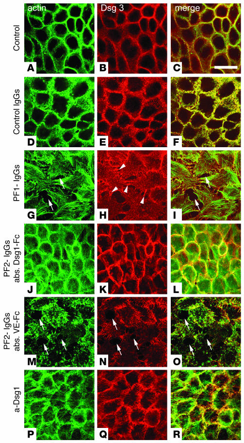
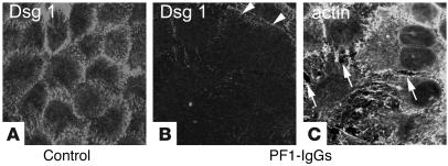
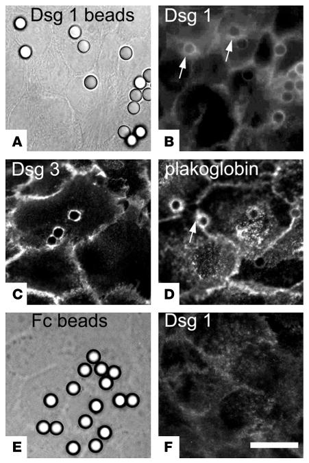
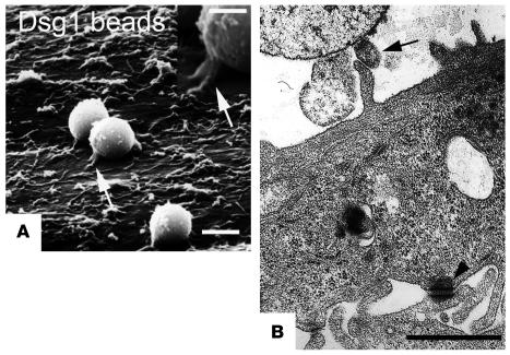
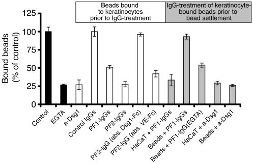
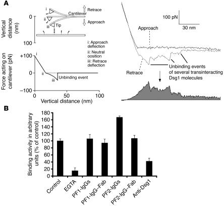
Autoantibodies against the epidermal desmosomal cadherins desmoglein 1 (Dsg1) and Dsg3 have been shown to cause severe to lethal skin blistering clinically defined as pemphigus foliaceus (PF) and pemphigus vulgaris (PV). It is unknown whether antibody-induced dissociation of keratinocytes is caused by direct inhibition of Dsg1 transinteraction or by secondary cellular responses. Here we show in an in vitro system that IgGs purified from PF patient sera caused cellular dissociation of cultured human keratinocytes as well as significant release of Dsg1-coated microbeads attached to Dsg-containing sites on the keratinocyte cellular surface. However, cell dissociation and bead release induced by PF-IgGs was not caused by direct steric hindrance of Dsg1 transinteraction, as demonstrated by single molecule atomic force measurements and by laser trapping of surface-bound Dsg1-coated microbeads. Rather, our experiments strongly indicate that PF-IgG-mediated dissociation events must involve autoantibody-triggered cellular signaling pathways, resulting in destabilization of Dsg1-based adhesive sites and desmosomes.


Figures

Figure 1
Figure 1
Characterization of PF-IgGs and HaCaT cells. Dot blot immunodetection demonstrating that the mouse monoclonal antibody directed against Dsg1 (i) as well as PF1-IgGs (iii) but not control IgGs (ii) detected the recombinant fusion protein consisting of the complete extracellular domain of Dsg1 and the Fc-portion of human IgGs (Dsg1-Fc). Both Dsg1 and Dsg3 were detected in HaCaT cell lysates by the monoclonal anti-Dsg1 (a-Dsg1) antibody (iv–vi) and the monoclonal a-Dsg3 antibody (vii), respectively. Contents of Dsg1 constantly increased from 3 days (iv) and 5 days (v) up to 7 days after plating (vi), the time when monolayers were used for experiments. The data shown is for 1 representative experiment out of 3.
Figure 2
Figure 2
PF-IgG–induced cell dissociation in HaCaT monolayers. HaCaT cells double-stained for F-actin using Alexa-phalloidin (A, D, G, J, M, and P) and Dsg3 (B, E, H, K, N, and Q). In controls, F-actin and the desmosomal cadherin Dsg3 were distributed along cell junctions (AC). Control IgGs (35 μg/ml, 24 hours) did not affect distribution of Dsg3 (E and F). In contrast, PF1-IgGs (35 μg/ml, 24 hours) induced intercellular gaps (arrows) best seen in the Alexa-phalloidin stain for F-actin (G and I). Note that Dsg3 is still present in cell processes spanning gaps (arrowheads in H). Following immunoabsorption of PF-IgGs by Dsg1-Fc–coated beads, PF2-IgGs (35 μg/ml, 24 hours) had no effect (JL) whereas large gaps (arrows) were induced by incubation with PF2-IgGs when control absorption was performed with beads coated with VE-cadherin–Fc (VE-Fc) (MO), indicating requirement of autoantibodies specific for Dsg1 for cell dissociation. Note that the inhibitory monoclonal antibody directed against the extracellular domain of Dsg1 did not induce gaps (PR). Scale bar: 40 μm for all panels (n = 5). abs., immunoabsorption.
Figure 3
Figure 3
PF-IgG–induced cell dissociation is accompanied by loss of Dsg1 staining. After incubation of HaCaT cells with PF-IgGs (35 μg/ml, 24 hours), monolayers were double-stained for Dsg1 (B) and F-actin using Alexa-phalloidin (C). Intercellular gaps are indicated by arrows. Dsg1 is located along cell borders (A) and becomes strongly reduced in response to PF-IgGs (arrowheads in B). Scale bar: 40 μm for all panels (n = 5).
Figure 4
Figure 4
Localization of Dsg1, Dsg3, and plakoglobin at cell-to-bead contacts. Dsg1-coated beads (AD) and Fc-coated beads (E and F) were immunostained following settlement on HaCaT cells for 30 minutes. Immunostaining for Dsg1 (B) showed localization of Dsg1 at the cell surface underneath most Dsg1-coated beads, which are visualized in A by corresponding phase contrast microscopic image. Note halo-like accumulation of cellular Dsg1 and plakoglobin around the bead attachment sites (arrows in B and D). Similarly, Dsg3 (C) was detected at cell-bead contacts. In contrast, no immunoreactivity for Dsg1 was found underneath Fc-coated beads visualized in a corresponding phase contrast figure (E and F). Scale bar: 10 μm for all panels (n = 5).
Figure 5
Figure 5
Characterization of cell-to-bead contacts induced by Dsg1-coated beads. Scanning electron microscopy (A) and transmission electron microscopy (B) were used to characterize contacts between HaCaT cells and Dsg1-coated beads formed after 30 minutes of settlement. Finger-like processes were present underneath beads with direct contact to HaCaT cells (arrows). These processes were similar to the desmosome-bearing processes found at contact sites between neighboring cells (arrowhead). Scale bars: 2.5 μm (A), 1.25 μm (A, inset); 1.0 μm (B).
Figure 6
Figure 6
Effect of PF-IgGs on binding of Dsg1-coated beads to HaCaT cells. Beads were allowed to settle on the surface of HaCaT cells for 30 minutes (control). Number of bound beads was reduced by simultaneous incubation of EGTA (5 mM, 30 minutes). Incubation of monolayers with attached beads for an additional 30 minutes with IgG fractions from 2 patients (PF1- and PF2-IgG, 35 μg/ml each) as well as with a monoclonal antibody (1:50) directed against the extracellular domain of human Dsg1 significantly reduced the number of bound beads (label in white box and white bars). Immunoabsorption using Dsg1-coated beads but not control absorption using VE-cadherin–labeled beads completely abolished the effect of PF-IgGs on bead adhesion. Preincubation of HaCaT cells with PF-IgGs prior to bead settlement also reduced bead binding (label in gray box and gray bars) whereas preincubation of beads with PF-IgGs did not inhibit bead binding. In contrast, the monoclonal Dsg1 antibody reduced bead binding also when applied for preincubation with beads, indicating a different mechanism underlying the reduction of Dsg1 adhesion (n = 6 for each condition).
Figure 7
Figure 7
Binding activity of Dsg1 in the presence or absence of PF-IgGs probed by AFM. (A) General working principle of force-distance cycles. Dsg1-Fc is covalently attached by PEG-linkers to the plate and cantilever tip of the AFM setup. Molecules are brought into contact by downward movement of the tip. During upward movement, a downward deflection of the cantilever will occur if plate- and tip-bound Dsg1 molecules undergo binding (left). Retrace and approach were subtracted, and the area below the resulting curve was integrated and taken as a measure for the average binding activity (right, gray). (B) Bar diagram shows binding activities of Dsg1 molecules in the presence or absence of antibodies. Binding activity was significantly reduced by incubation with EGTA (5 mM, 30 minutes) or the monoclonal antibody directed against Dsg1 (1:50). However, PF-IgGs (35 μg/ml, 30 min) did not reduce binding events in this cell-free system. Fab fragments from the PF-IgG fraction have been applied to rule out possible cross-linking effects of the PF-IgGs and were found not to reduce Dsg1-mediated binding (n = 4 for each condition).

Republished in

References

    1. Stanley JR, Yaar M, Hawley-Nelson P, Katz SI. Pemphigus antibodies identify a cell surface glycoprotein synthesized by human and mouse keratinocytes. J. Clin. Invest. 1982;70:281–288. - PMC - PubMed
    1. Stanley JR, Koulu L, Thivolet C. Distinction between epidermal antigens binding pemphigus vulgaris and pemphigus foliaceus autoantibodies. J. Clin. Invest. 1984;74:313–320. - PMC - PubMed
    1. Amagai M, Karpati S, Prussick R, Klaus-Kovtun V, Stanley JR. Autoantibodies against the amino-terminal cadherin-like binding domain of pemphigus vulgaris antigen are pathogenic. J. Clin. Invest. 1992;90:919–926. - PMC - PubMed
    1. Amagai M, Hashimoto T, Green KJ, Shimizu N, Nishikawa T. Antigen-specific immunoadsorption of pathogenic autoantibodies in pemphigus foliaceus. J. Invest. Dermatol. 1995;104:895–901. - PubMed
    1. Nollet F, Kools P, van Roy F. Phylogenetic analysis of the cadherin superfamily allows identification of six major subfamilies besides several solitary members. J. Mol. Biol. 2000;299:551–572. - PubMed

Publication types

MeSH terms