

A variant of SCID with specific immune responses and predominance of gamma delta T cells
- PMID: 16211094
- PMCID: PMC1242191
- DOI: 10.1172/JCI25221
A variant of SCID with specific immune responses and predominance of gamma delta T cells
Abstract
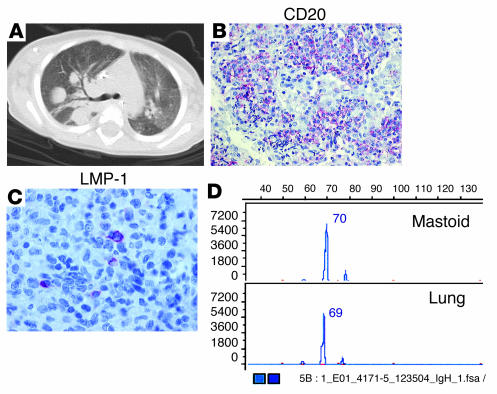
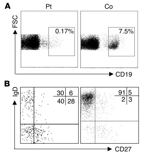
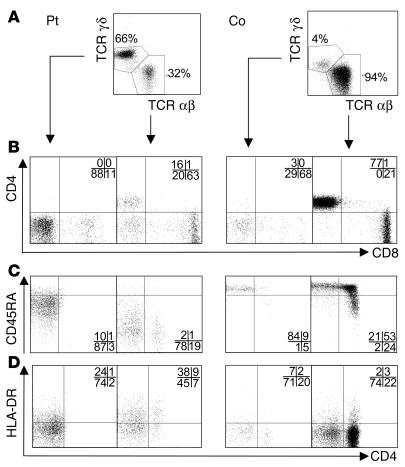
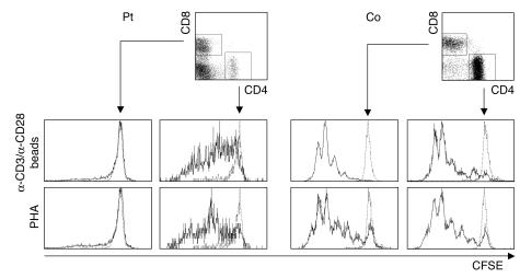
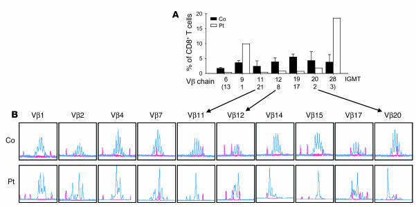
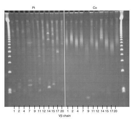
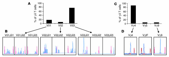
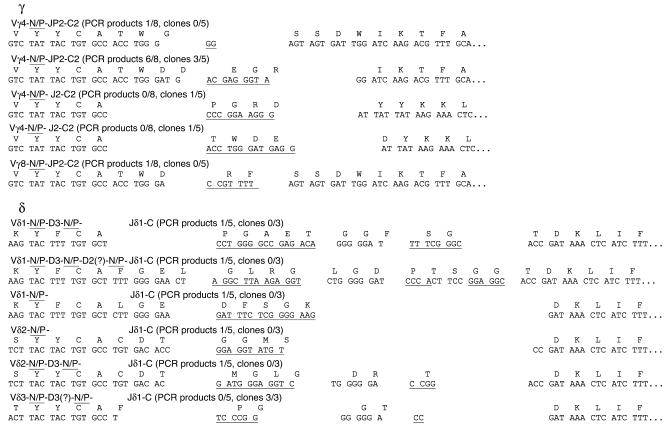
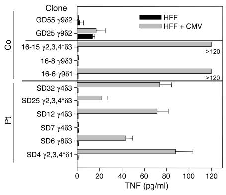
We describe here a patient with a clinical and molecular diagnosis of recombinase activating gene 1-deficient (RAG1-deficient) SCID, who produced specific antibodies despite minimal B cell numbers. Memory B cells were detected and antibodies were produced not only against some vaccines and infections, but also against autoantigens. The patient had severely reduced levels of oligoclonal T cells expressing the alphabeta TCR but surprisingly normal numbers of T cells expressing the gammadelta TCR. Analysis at a clonal level and TCR complementarity-determining region-3 spectratyping for gammadelta T cells revealed a diversified oligoclonal repertoire with predominance of cells expressing a gamma4-delta3 TCR. Several gammadelta T cell clones displayed reactivity against CMV-infected cells. These observations are compatible with 2 non-mutually exclusive explanations for the gammadelta T cell predominance: a developmental advantage and infection-triggered, antigen-driven peripheral expansion. The patient carried the homozygous hypomorphic R561H RAG1 mutation leading to reduced V(D)J recombination but lacked all clinical features characteristic of Omenn syndrome. This report describes a new phenotype of RAG deficiency and shows that the ability to form specific antibodies does not exclude the diagnosis of SCID.
Figures
Similar articles
-
Expression of MHC class I receptors confers functional intraclonal heterogeneity to a reactive expansion of gammadelta T cells.Eur J Immunol. 2005 Jun;35(6):1896-905. doi: 10.1002/eji.200425837. Eur J Immunol. 2005. PMID: 15864777
-
A novel immunodeficiency associated with hypomorphic RAG1 mutations and CMV infection.J Clin Invest. 2005 Nov;115(11):3291-9. doi: 10.1172/JCI25178. J Clin Invest. 2005. PMID: 16276422 Free PMC article.
-
More than just SCID--the phenotypic range of combined immunodeficiencies associated with mutations in the recombinase activating genes (RAG) 1 and 2.Clin Immunol. 2010 May;135(2):183-92. doi: 10.1016/j.clim.2010.01.013. Epub 2010 Feb 20. Clin Immunol. 2010. PMID: 20172764 Review.
-
Alphabeta T-cell receptor engineered gammadelta T cells mediate effective antileukemic reactivity.Cancer Res. 2006 Mar 15;66(6):3331-7. doi: 10.1158/0008-5472.CAN-05-4190. Cancer Res. 2006. PMID: 16540688
-
Gammadelta T cells: functional plasticity and heterogeneity.Nat Rev Immunol. 2002 May;2(5):336-45. doi: 10.1038/nri797. Nat Rev Immunol. 2002. PMID: 12033739 Review.
Cited by
-
TCR-dependent sensitization of human γδ T cells to non-myeloid IL-18 in cytomegalovirus and tumor stress surveillance.Oncoimmunology. 2015 Feb 3;4(5):e1003011. doi: 10.1080/2162402X.2014.1003011. eCollection 2015 May. Oncoimmunology. 2015. PMID: 26155394 Free PMC article.
-
Outcomes and Treatment Strategies for Autoimmunity and Hyperinflammation in Patients with RAG Deficiency.J Allergy Clin Immunol Pract. 2019 Jul-Aug;7(6):1970-1985.e4. doi: 10.1016/j.jaip.2019.02.038. Epub 2019 Mar 12. J Allergy Clin Immunol Pract. 2019. PMID: 30877075 Free PMC article.
-
DCLRE1C (ARTEMIS) mutations causing phenotypes ranging from atypical severe combined immunodeficiency to mere antibody deficiency.Hum Mol Genet. 2015 Dec 20;24(25):7361-72. doi: 10.1093/hmg/ddv437. Epub 2015 Oct 16. Hum Mol Genet. 2015. PMID: 26476407 Free PMC article.
-
Natural Killer Cells from Patients with Recombinase-Activating Gene and Non-Homologous End Joining Gene Defects Comprise a Higher Frequency of CD56bright NKG2A+++ Cells, and Yet Display Increased Degranulation and Higher Perforin Content.Front Immunol. 2017 Jul 17;8:798. doi: 10.3389/fimmu.2017.00798. eCollection 2017. Front Immunol. 2017. PMID: 28769923 Free PMC article.
-
γδ T Cell-Mediated Immunity to Cytomegalovirus Infection.Front Immunol. 2017 Feb 9;8:105. doi: 10.3389/fimmu.2017.00105. eCollection 2017. Front Immunol. 2017. PMID: 28232834 Free PMC article. Review.
References
-
- Buckley RH. Molecular defects in human severe combined immunodeficiency and approaches to immune reconstitution. Annu. Rev. Immunol. 2004;22:625–655. - PubMed
-
- Buckley RH, et al. Human severe combined immunodeficiency: genetic, phenotypic, and functional diversity in one hundred eight infants. J. Pediatr. 1997;130:378–387. - PubMed
-
- Omenn GS. Familial reticuloendotheliosis with eosinophilia. N. Engl. J. Med. 1965;273:427–432. - PubMed
-
- Villa A, et al. Partial V(D)J recombination activity leads to Omenn syndrome. Cell. 1998;93:885–896. - PubMed
-
- Villa A, et al. V(D)J recombination defects in lymphocytes due to RAG mutations: severe immunodeficiency with a spectrum of clinical presentations. Blood. 2001;97:81–88. - PubMed
Publication types
MeSH terms
Substances
LinkOut - more resources
Full Text Sources
Other Literature Sources
