Review
. 2005 Dec;26(16):1752-80.
doi: 10.1002/jcc.20292.

Integrated Modeling Program, Applied Chemical Theory (IMPACT)

Affiliations
Review

Integrated Modeling Program, Applied Chemical Theory (IMPACT)

Jay L Banks et al. J Comput Chem. 2005 Dec.

Abstract

We provide an overview of the IMPACT molecular mechanics program with an emphasis on recent developments and a description of its current functionality. With respect to core molecular mechanics technologies we include a status report for the fixed charge and polarizable force fields that can be used with the program and illustrate how the force fields, when used together with new atom typing and parameter assignment modules, have greatly expanded the coverage of organic compounds and medicinally relevant ligands. As we discuss in this review, explicit solvent simulations have been used to guide our design of implicit solvent models based on the generalized Born framework and a novel nonpolar estimator that have recently been incorporated into the program. With IMPACT it is possible to use several different advanced conformational sampling algorithms based on combining features of molecular dynamics and Monte Carlo simulations. The program includes two specialized molecular mechanics modules: Glide, a high-throughput docking program, and QSite, a mixed quantum mechanics/molecular mechanics module. These modules employ the IMPACT infrastructure as a starting point for the construction of the protein model and assignment of molecular mechanics parameters, but have then been developed to meet specialized objectives with respect to sampling and the energy function.


Figures

Figure 1
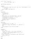
Figure 1
A sample IMPACT input file.
Figure 2
Figure 2
The potential of mean force at 298 K of the capped C-terminal peptide from protein G with respect to the second (PC2) and third (PC3) principal components (PC3 corresponds approximately to the end-to-end distance), using the OPLS-AA/AGBNP potential with additional dielectric screening of charged side chains. The PMF is calculated from T-WHAM analysis of the ensemble of structures generated by all RXMD replicas with temperatures from 270 to 700 K. The energy is in units of kcal/mol. The low free energy region to the left correspond to α-helical conformations. The wider free energy basin to the right correspond to β-hairpin conformations. The black and white paths correspond to the upper and lower α-helical to β-hairpin conversion mechanisms shown in Figure 3, respectively. [Color figure can be viewed in the online issue, which is available at www.interscience.wiley.com.]
Figure 3
Figure 3
Two possible pathways for the interconversion of an α-helix into a β-hairpin. Backbone trace is shown in blue, and the hydrophobic core residues (W43, Y45, F52, and V54) side chains are shown in gold. The upper path corresponds to unraveling of the helix at both ends and formation of a β-turn from a residual turn of α-helix. The lower path corresponds to unraveling of one end of the helix, which loops back.
Figure 4
Figure 4
The open ribose-free crystal structure of RBP (PDB id 1urp, left) and the closed ribose-bound crystal structure of RBP (PDB id 2dri, right). Ribose (D-ribopyranose form) is shown in ball-and-stick representation. The N-terminal domain of RBP is shown in blue and the C-terminal domain is shown in red. Three strands connecting the N-terminal domain to the C-terminal domain form the hinge region shown in green. The conformational change from the open (a) to the closed (b) conformation consists of a rotation of the C-terminal domain (red) toward the N-terminal domain (blue) around the axis perpendicular to the page centered on the hinge region (bending), followed by a rotation towards the viewer of the C-terminal domain around the axis longitudinal to the hinge region and parallel to the page (twisting). This figure has been generated using the program MOLSCRIPT.
Figure 5
Figure 5
Calculated population distribution of (a) ribose-free and (b) ribose-bound RBP as a function of the interdomain bending angle θ and twisting angle φ (see Fig. 4). Contours are drawn at 0.08 (green), 0.06 (blue), 0.04 (magenta), 0.03 (sky blue), 0.02 (yellow), 0.01 (brown), 0.001 (red), and 0.0001(gray) relative populations. The crosses indicate the (θ, φ) coordinates of the crystal structures of, with increasing angle θ, the closed ribose-bound (2dri) and open ribose-free (1urp) conformations of RBP, and open ribose-free conformations of a single-point mutant of RBP (1ba2). The angle θ is defined as the angle formed between the centers of mass of the C- and N-terminal domains and the center of mass of the hinge region. The angle φ is defined as the dihedral angle formed by the center of mass of the N-terminal domain, the center of mass of the residues on the N-terminal domain side of each of the three hinge strands, the center of mass of the corresponding residues on the C-terminal domain side of the three hinge strands, and the center of mass of the C-terminal domain. Angles are expressed in degrees. Highly populated conformations generally correspond to experimental crystal conformations. In agreement with experiments, the predicted population of the closed conformation (θ ≈ 110°, φ ≈ 50°) in the presence of ribose (b) is larger than in the absence of ribose (a). Similarly, the population of open conformations (θ >115°) is larger in the absence of ribose. The population peak at (θ ≈ 122°, φ ≈ 95°) in the presence of ribose (b) corresponds to a partially open conformation not yet observed experimentally postulated to play a role in the mechanism of ribose transport.
Figure 6
Figure 6
RMSD from the native of the docked ligands using FF Dock and QM Dock.
Figure 7
Figure 7
RMSD from the native of the docked ligands using FF Dock and QM Dock with the “SOF” algorithm.

References

    1. McCammon JA, Gelin BR, Karplus M. Nature. 1977;267:585. - PubMed
    1. Karplus M. Biopolymers. 2003;68:350. - PubMed
    1. Levy RM, Bassolino D, Kitchen DB, Pardi A. Biochemistry. 1989;28:9361. - PubMed
    1. Fan P, Kominos D, Kitchen DB, Levy RM, Baum J. Chem Phys. 1991;158:295.
    1. Kitchen DB, Hirata F, Kofke DA, Westbrook JD, Yarmush M, Levy RM. J Comput Chem. 1990;11:1169.

Publication types