. 2005 Oct 7;123(1):89-103.
doi: 10.1016/j.cell.2005.07.032.

Metabolic regulation of oocyte cell death through the CaMKII-mediated phosphorylation of caspase-2

Affiliations

Metabolic regulation of oocyte cell death through the CaMKII-mediated phosphorylation of caspase-2

Leta K Nutt et al. Cell. .

Abstract

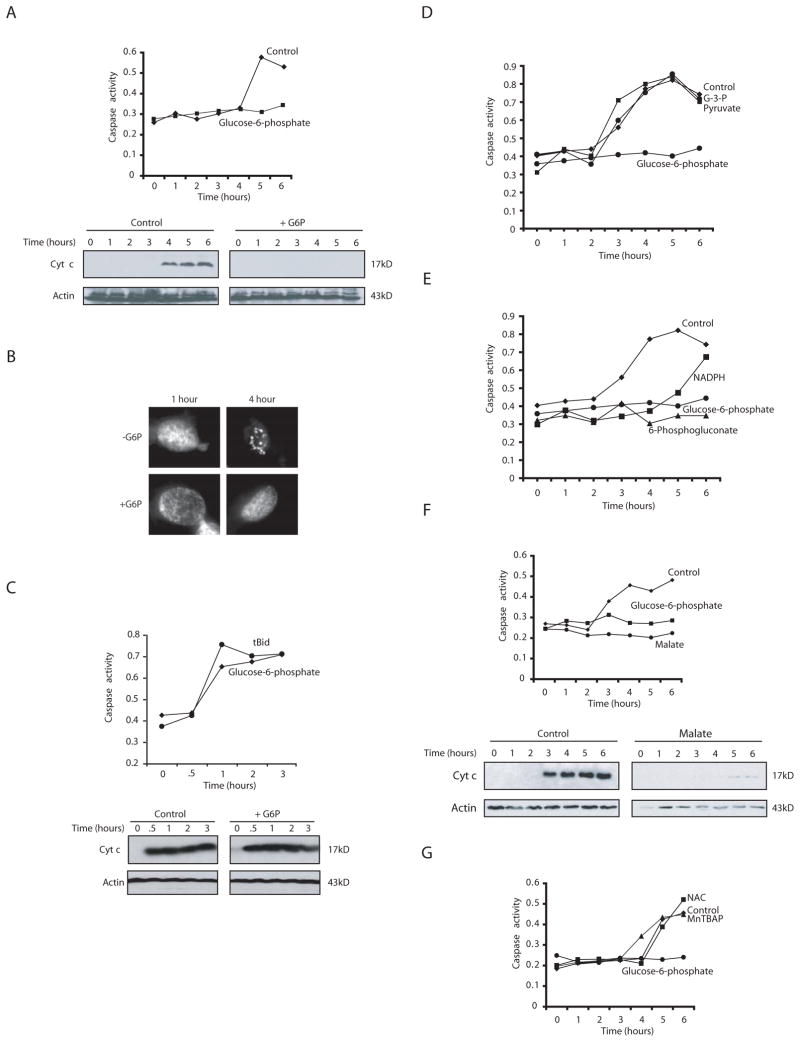
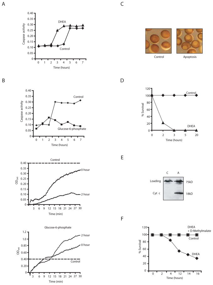
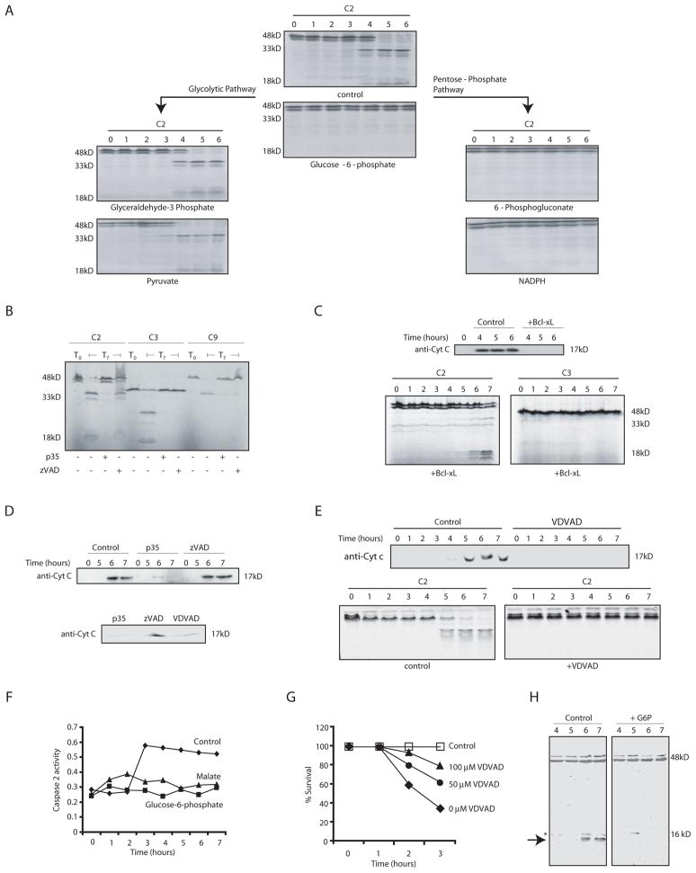
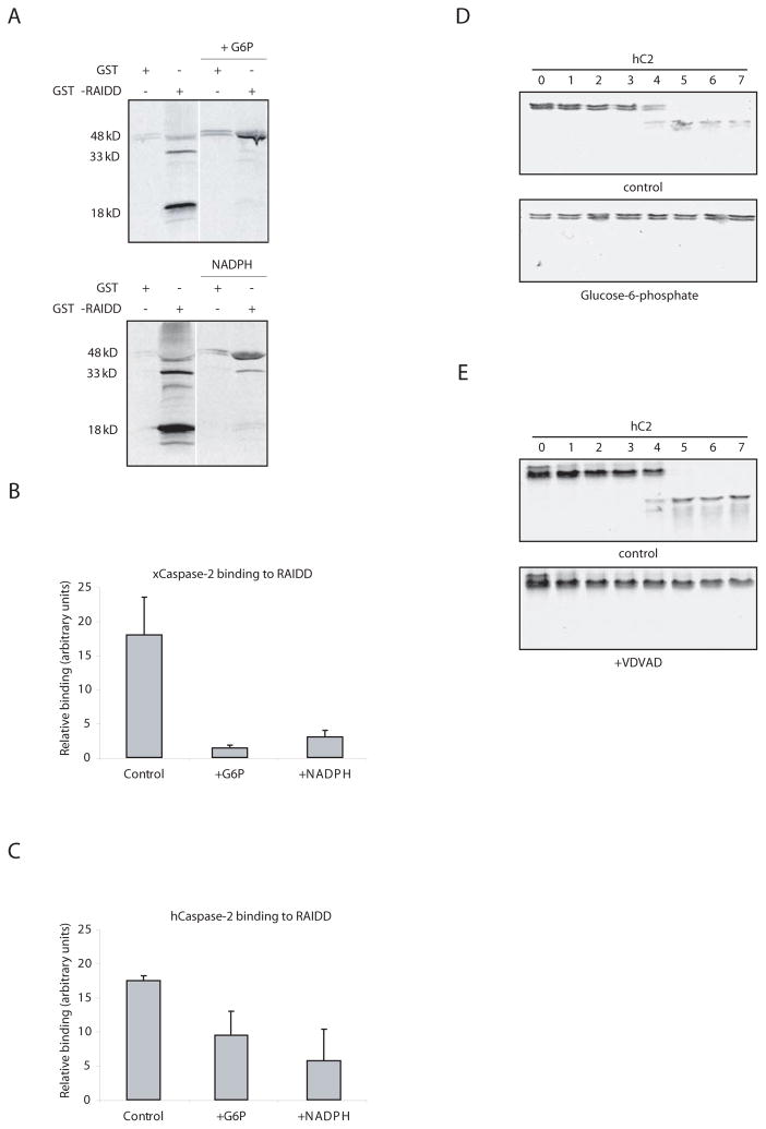
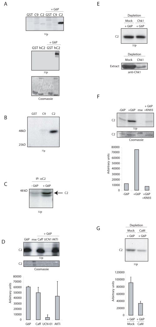
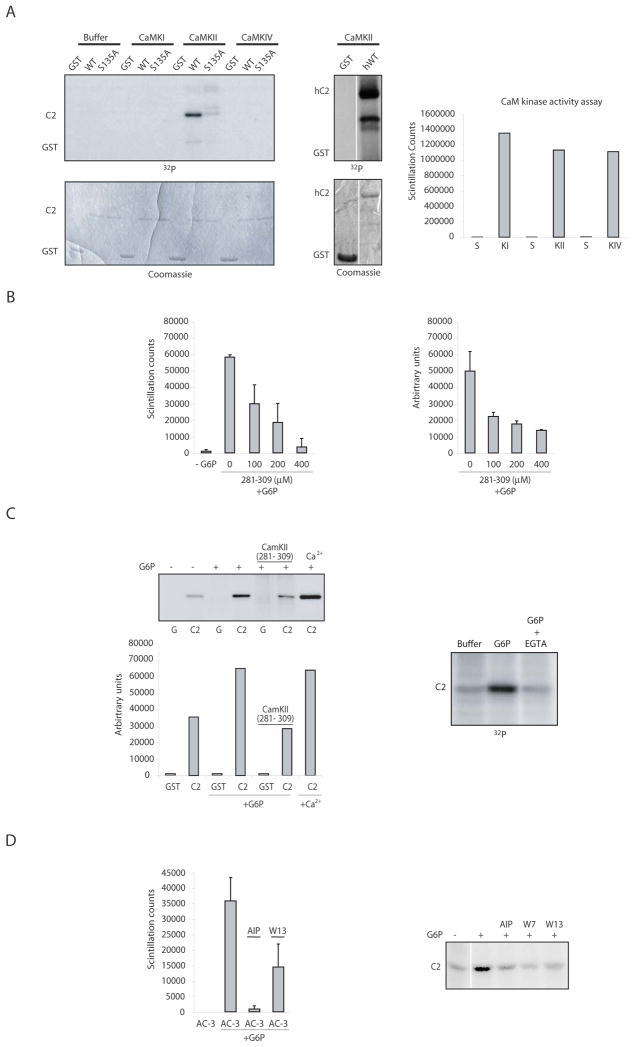
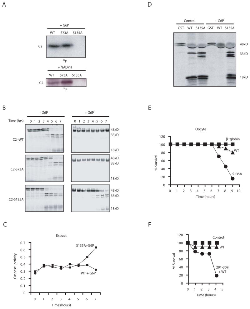
Vertebrate female reproduction is limited by the oocyte stockpiles acquired during embryonic development. These are gradually depleted over the organism's lifetime through the process of apoptosis. The timer that triggers this cell death is yet to be identified. We used the Xenopus egg/oocyte system to examine the hypothesis that nutrient stores can regulate oocyte viability. We show that pentose-phosphate-pathway generation of NADPH is critical for oocyte survival and that the target of this regulation is caspase-2, previously shown to be required for oocyte death in mice. Pentose-phosphate-pathway-mediated inhibition of cell death was due to the inhibitory phosphorylation of caspase-2 by calcium/calmodulin-dependent protein kinase II (CaMKII). These data suggest that exhaustion of oocyte nutrients, resulting in an inability to generate NADPH, may contribute to ooctye apoptosis. These data also provide unexpected links between oocyte metabolism, CaMKII, and caspase-2.


Figures

Fig. 1
Fig. 1. NADPH is a potent inhibitor of egg apoptosis
A) Xenopus egg extracts supplemented with G6P or buffer were analyzed for caspase 3 activity at various time points using the caspase substrate Ac-DEVD-pNA. Substrate cleavage was measured spectrophotometrically at 405nm. Cytochrome c release was measured in parallel by filtering extract through a 0.1μm microfilter. The filtrate, lacking mitochondria, was analyzed by SDS-PAGE and immunoblotting with anti-cytochrome c antibody. Blots were re-probed with anti-Actin antibody. B) Extracts treated as in A were supplemented with sperm nuclei and stained with Hoechst dye to detect DNA by fluorescence microscopy. Shown are representative nuclei at the indicated times. C) Truncated Bid (tBid) was added to extract treated with G6P. Caspase 3 activity and cytochrome c release were measured as in (A). D) Buffer, glyceraldehyde-3-phosphate (G-3-P), pyruvate, or G6P were added to extract and caspase 3 activity was measured. E) Pentose phosphate intermediates (NADPH, 6-phosphogluconate) or G6P were added to extract and caspase 3 activity measured. F) Malate was added to extracts and caspase 3 activity and cytochrome c release measured. G) A superoxide dismutase mimetic Mn(III)tetrakis(4–Benzoic acid)porphyrin Chloride (MnTBAP) or a precursor to glutathione, N-Acetyl-L-cysteine (NAC) was added to extracts and caspase activity was measured.
Fig. 2
Fig. 2. Inhibition of G6P dehydrogenase induces apoptosis
A) Extracts supplemented with either dehydroisoandrosterone (DHEA) or buffer were analyzed for caspase 3 activity. Shown is a representative experiment repeated on 3 separate batches of oocytes with similar results. B) (upper): Extract was incubated at room temp and assayed for caspase activity. (middle): Samples withdrawn at 0 and 2 h of incubation were assayed for G6P levels by monitoring NADPH production over time (absorbance at 340 nm) in the presence of excess G6P dehydrogenase and NADP. (lower): Addition of excess G6P maintained NADPH production at all times tested. Dotted line indicates the fact that the scales are different in the extracts +/− G6P as NADPH was produced at much higher rates in the presence of excess G6P. C) Oocytes treated with DHEA or buffer are shown in representative micrographs. D) Percent survival of oocytes treated with buffer or DHEA. E) Cytochrome c release was measured in parallel by filtering aliquots of lysed oocytes through a 0.1μm microfilter and analyzing the filtrate by anti-cytochrome c immunoblotting. F) Percent survival of oocytes treated with buffer, DHEA or DHEA and cell permeable D-methyl malate.
Fig. 3
Fig. 3. Pentose phosphate metabolites act upstream of cytochrome c release
A) Egg extract was incubated with metabolic substrates and samples were analyzed for processing of in vitro translated caspase 2. B) Extract was incubated at room temp in the presence of caspase inhibitors (zVAD, 100 μM, VDVAD 100μM, or p35) and radiolabeled pro-caspases. Samples from time 0 (T0) and 7 hours (T7) were analyzed by SDS-page for processing of in vitro translated caspases 2 (C2), 3 (C3), and 9 (C9). C) Extract was incubated at room temp +/− Bcl-xL, and samples were analyzed every hour for in vitro translated C2 and C3 processing by SDS-PAGE and for cytochrome c release. D) (upper panel): Egg extract was incubated at room temp and treated with or without p35 and zVAD-fmk. Samples were taken every hour, mitochondria removed by filtration and the remaining cytosol immunoblotted with anti-cytochrome c antibody. (lower panel) same as above except a 7 h timepoint is shown also in the presence of VDVAD. E) (Upper panel): Samples were treated as in D, but in the presence of VDVAD (lower panel): Processing of radiolabeled caspase 2 was monitored +/− VDVAD. F) Cleavage of VDVAD-pNA was monitored in extracts containing buffer, malate or G6P. G) Oocytes were treated with DHEA as in 2A, with or without prior soaking in buffer containing the indicated concentrations of VDVAD. H) Cleavage of endogenous caspase 2 was monitored +/− G6P by anti-caspase 2 immunoblotting using a mix of C20 and H119 antibodies from Santa Cruz, which recognize only the full length 48 kD protein and the 12 kD cleavage fragment, indicated by the arrow. The asterisk indicates a background band present in all lanes.
Fig. 4
Fig. 4. Anti-apoptotic effects of NADPH are exerted at the level of caspase 2
A) GST-RAIDD or GST bound to glutathione sepharose was dipped into extract +/− NADPH or G6P in the presence of radiolabeled pro-caspase 2. Beads were retrieved by centrifugation, boiled in SDS sample buffer and resolved by SDS-PAGE for autoradiography. Pro-caspase 2 not bound to beads was also unprocessed in the presence of G6P or NADPH (data not shown). B) GST-RAIDD sepharose was dipped into extracts with in vitro translated caspase 2 in the presence and absence of NADPH. Beads were washed extensively and radiolabeled caspase 2 binding was measured by phosphoimager. C) The same experiment in B was repeated using in vitro translated human, rather than Xenopus, caspase 2. D) Processing of radiolabeled human pro-caspase 2 supplemented into egg extracts was monitored in the presence and absence of G6P. E) The same as in D, but +/− VDVAD.
Fig. 5
Fig. 5. G6P/NADPH induces phosphorylation and inactivation of caspase 2
A) (upper) The GST-prodomain of Xenopus caspase 2 (C2) or 9 (C9) was bound to glutathione sepharose and dipped into cytosolic extracts +/− G6P, and γ-32P-ATP. Bead-bound proteins were washed and analyzed by SDS-PAGE and phosphorimaging. (lower): Similar results were obtained with the human caspase 2 prodomain. B) The experiment in A) was repeated minus G6P and over-exposed to detect basal caspase 2 phosphorylation. C) Anti-Xenopus caspase 2 prepared against a C-terminal 20 amino acid peptide or preimmune antibodies were bound to protein A beads and dipped into extract +/− G6P and γ-32P-ATP. Samples were resolved by SDS-PAGE and detected by autoradiography. D) The experiment in A) was repeated with 5 mM Caffeine, UCN-01 (1 μM), or Akt inhibitor (20 μM). Sepharose-bound GST proteins were washed and analyzed by SDS-PAGE and quantitated by phosphorimaging. E) Egg extracts were depleted with anti-Chk1 or control antibody and incubated with G6P. Samples were washed and analyzed by SDS-PAGE and autoradiography (upper panel) or by anti-Chk1 immunoblotting (lower panel). F) The experiment in A) was repeated with 5μM KN-93. Sepharose-bound GST proteins were washed and analyzed by SDS-PAGE and quantitated by phosphorimaging. G) Egg extracts were depleted with calmodulin sepharose or control sepharose and then incubated with G6P. Caspase 2 prodomain phosphorylation was monitored as in A.
Fig 6
Fig 6. CaMKII phosphorylates the prodomain of caspase 2
A) (left): In vitro kinase assays were performed with either buffer, CaMKI, CaMKII or CaMKIV and the GST-fused prodomain of caspase 2 (WT or S135A). Prodomain phosphorylation was analyzed by SDS-PAGE, and the gel was stained with coomassie blue to show equal loading. (middle): The same assay was performed using CaMKII and the human caspase 2 prodomain. (right): The activity of each kinase was tested against its cognate peptide substrate and compared to substrate (S) alone in the absence of kinase (right panel). B) (Left): Extract was incubated +/− G6P, γ-32P-ATP, and the peptide substrate Syntide-2 (Syn2) +/− various doses of a CaMKII peptide inhibitor (CaMKII 281–309). Substrate trapped on filter paper was washed extensively and scintillation counted. (right): The same extracts were tested using GST (G) or GST-caspase 2 prodomain (C2) as substrates by the same protocol as in Fig. 5B (right panel shows quantiation of GST-prodomain phosphorylation from 3 such gels exposed to the indicated inhibitor doses). C) (left upper panel): A represenative gel for the experiment shown in the right hand panel of B, with quantitation of the gel, below. (right): The assay in Fig. 5B was repeated in the presence of EGTA. D) (right): Using the same conditions as in B), another CaMKII ινηιβιτoρ (5 μM AIP) was tested using its peptide substrate AC- 3 (left) or the caspase 2 prodomain. (right): Calmodulin inhibitors (10 μM W13 and 10 μM W7) were tested in the same manner.
Fig. 7
Fig. 7. Mutation of S135 of caspase 2 to Ala abrogates the protective effects of NADPH
A) The WT, S73A and S135A caspase 2 prodomains fused to GST were bound to glutathione sepharose and dipped into cytosolic extracts +/− G6P, +/− NADPH, and γ-32P-ATP. Sepharose-bound proteins were washed, eluted with sample buffer and resolved by SDS-PAGE. B) Extracts supplemented with WT, S135A, or S73A radiolabeled pro-caspase 2 were incubated at room temp and treated with or without G6P. Samples were taken at the indicated times and analyzed by SDS-PAGE. C) Pro-Caspase 2 S135A or WT mRNA and G6P were added to a translationally competent egg extract and samples were taken to measure cleavage of Ac-DEVD-pNA. D) GST-RAIDD was added to extracts with in vitro translated WT or S135A pro-caspase 2 +/− G6P for 1 h. Samples were taken to measure caspase 2 processing by SDS-PAGE. E) Oocytes were injected with mRNA encoding WT or S135A pro-caspase 2, or β-globin. Cell death was quantitated as in Fig. 2. Shown is a representative experiment repeated on 3 oocyte batches with similar results. F) Oocytes were injected with the 281–309 CaMKII inhibitory peptide and subsequently injected with WT pro-caspase 2-encoding mRNA and monitored as in E.

Comment in

Similar articles

Cited by

References

    1. Bagowski CP, Xiong W, Ferrell JE., Jr c-Jun N-terminal kinase activation in Xenopus laevis eggs and embryos. A possible non-genomic role for the JNK signaling pathway. J Biol Chem. 2001;276:1459–1465. - PubMed
    1. Bergeron L, Perez GI, Macdonald G, Shi L, Sun Y, Jurisicova A, Varmuza S, Latham KE, Flaws JA, Salter JC, et al. Defects in regulation of apoptosis in caspase-2-deficient mice. Genes Dev. 1998;12:1304–1314. - PMC - PubMed
    1. Bhuyan AK, Varshney A, Mathew MK. Resting membrane potential as a marker of apoptosis: studies on Xenopus oocytes microinjected with cytochrome c. Cell Death Differ. 2001;8:63–69. - PubMed
    1. Blitzer RD, Connor JH, Brown GP, Wong T, Shenolikar S, Iyengar R, Landau EM. Gating of CaMKII by cAMP-regulated protein phosphatase activity during LTP. Science. 1998;280:1940–1942. - PubMed
    1. Braun T, Dar S, Vorobiov D, Lindenboim L, Dascal N, Stein R. Expression of Bcl-x(S) in Xenopus oocytes induces BH3-dependent and caspase-dependent cytochrome c release and apoptosis. Mol Cancer Res. 2003;1:186–194. - PubMed

Publication types

MeSH terms