

Constitutive activation of prosurvival signaling in alveolar mesenchymal cells isolated from patients with nonresolving acute respiratory distress syndrome
- PMID: 16214815
- PMCID: PMC1382273
- DOI: 10.1152/ajplung.00276.2005
Constitutive activation of prosurvival signaling in alveolar mesenchymal cells isolated from patients with nonresolving acute respiratory distress syndrome
Abstract
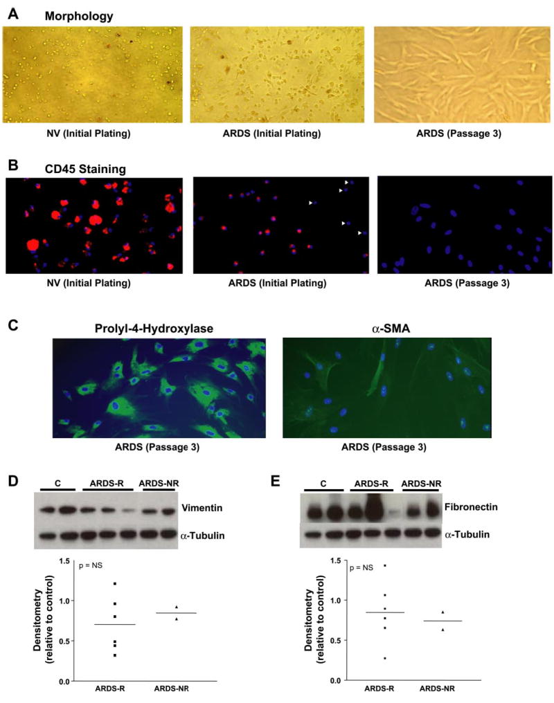
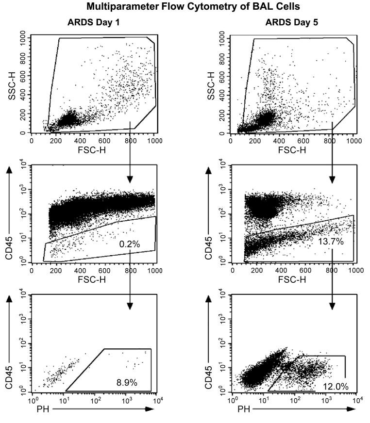
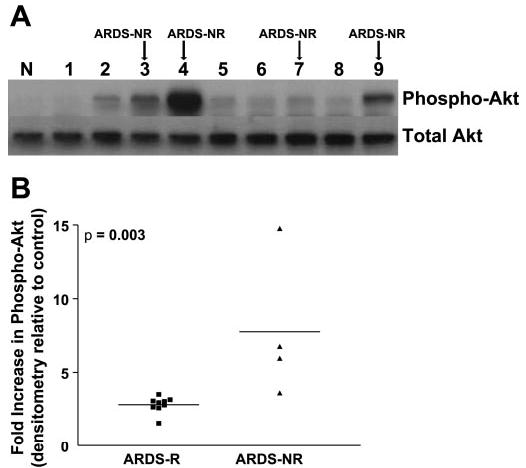
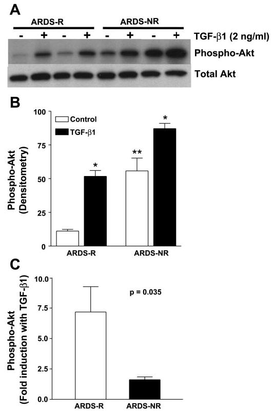
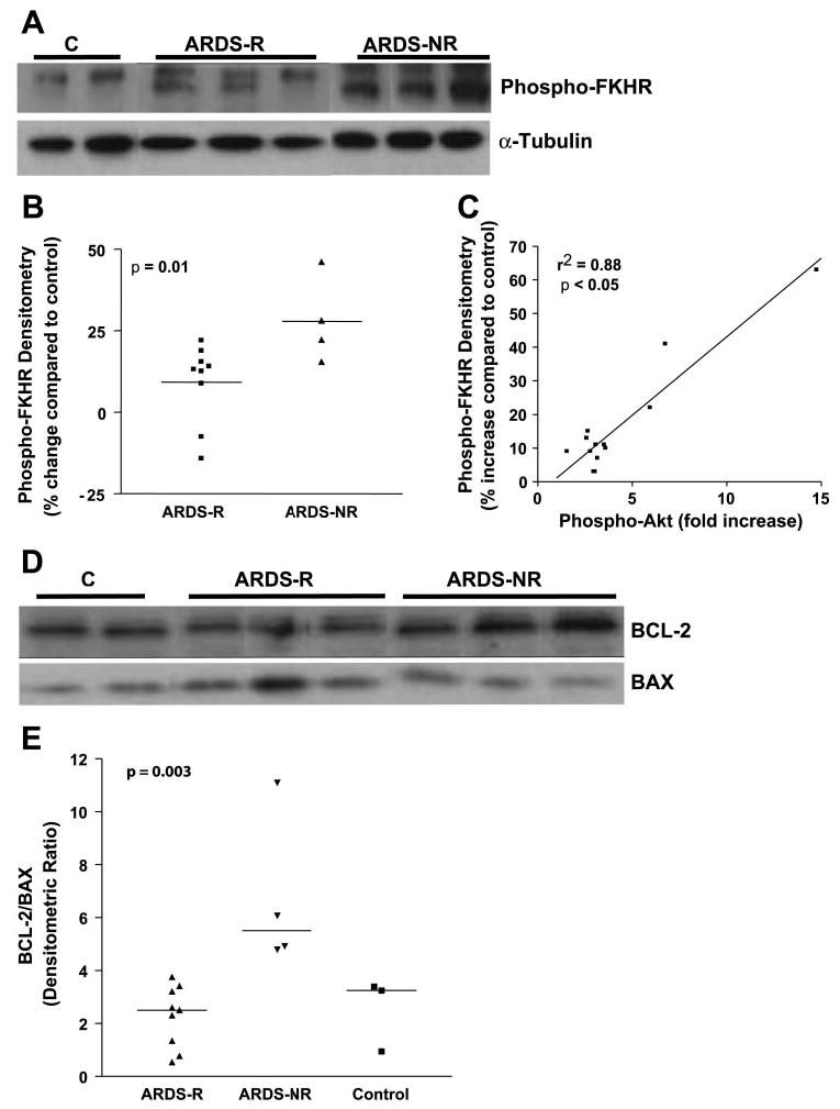
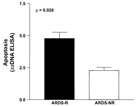
Acute respiratory distress syndrome (ARDS) is a clinical syndrome characterized by stereotypic host inflammatory and repair cellular responses; however, mechanisms regulating the resolution of ARDS are poorly understood. Here, we report the isolation and characterization of a novel population of mesenchymal cells from the alveolar space of ARDS patients via fiber-optic bronchoscopy with bronchoalveolar lavage (BAL). BAL was performed on 17 patients during the course of ARDS. Immunofluorescence staining and multiparameter flow cytometric analysis defined a population of alveolar mesenchymal cells (AMCs) that are CD45-/prolyl-4-hydroxylase+/alpha-smooth muscle actin+/-. AMCs proliferated in ex vivo cell culture for multiple passages; early passage (3-5) cells were subsequently analyzed in 13 patients. AMCs isolated from patients with persistent or nonresolving ARDS (ARDS-NR, n = 4) demonstrate enhanced constitutive activation of prosurvival signaling pathways involving PKB/Akt, FKHR, and BCL-2 family proteins compared with AMCs from patients with resolving ARDS (ARDS-R, n = 9). Exogenous transforming growth factor-beta1 markedly induces PKB/Akt activation in AMCs from ARDS-R. ARDS-NR cells are more resistant to serum deprivation-induced apoptosis compared with ARDS-R. This study identifies a novel population of mesenchymal cells that can be isolated from the alveolar spaces of ARDS patients. AMCs in patients with ARDS-NR acquire an activational profile characterized by enhanced prosurvival signaling and an antiapoptotic phenotype. These findings support the concept that apoptosis of mesenchymal cells may be an essential component of normal repair and resolution of ARDS and suggest that dysregulation of this process may contribute to persistent ARDS.
Figures
Similar articles
-
Alveolar fibroblasts in acute lung injury: biological behaviour and clinical relevance.Eur Respir J. 2010 Jun;35(6):1312-21. doi: 10.1183/09031936.00074709. Epub 2009 Oct 19. Eur Respir J. 2010. PMID: 19840966
-
Early infectious acute respiratory distress syndrome is characterized by activation and proliferation of alveolar T-cells.Eur J Clin Microbiol Infect Dis. 2015 Jun;34(6):1111-8. doi: 10.1007/s10096-015-2333-x. Epub 2015 Feb 5. Eur J Clin Microbiol Infect Dis. 2015. PMID: 25652606
-
Keratinocyte growth factor expression is suppressed in early acute lung injury/acute respiratory distress syndrome by smad and c-Abl pathways.Crit Care Med. 2009 May;37(5):1678-84. doi: 10.1097/CCM.0b013e31819fc81a. Crit Care Med. 2009. PMID: 19325470
-
Bronchoalveolar lavage in patients with the adult respiratory distress syndrome.Clin Chest Med. 1985 Sep;6(3):459-71. Clin Chest Med. 1985. PMID: 3907947 Review.
-
The Complex Immune Cell Composition and Cellular Interaction in the Alveolar Compartment of Patients with Acute Respiratory Distress Syndrome.Am J Respir Cell Mol Biol. 2025 Mar;72(3):233-243. doi: 10.1165/rcmb.2024-0176TR. Am J Respir Cell Mol Biol. 2025. PMID: 39383858 Free PMC article. Review.
Cited by
-
Prostaglandin E(2) induces fibroblast apoptosis by modulating multiple survival pathways.FASEB J. 2009 Dec;23(12):4317-26. doi: 10.1096/fj.08-128801. Epub 2009 Aug 11. FASEB J. 2009. PMID: 19671668 Free PMC article.
-
Advances in mechanisms of repair and remodelling in acute lung injury.Intensive Care Med. 2008 Apr;34(4):619-30. doi: 10.1007/s00134-007-0963-x. Epub 2008 Feb 9. Intensive Care Med. 2008. PMID: 18264692 Review.
-
Early activation of pro-fibrotic WNT5A in sepsis-induced acute lung injury.Crit Care. 2014 Oct 21;18(5):568. doi: 10.1186/s13054-014-0568-z. Crit Care. 2014. PMID: 25331176 Free PMC article.
-
Inhibition of antiapoptotic BCL-2 proteins with ABT-263 induces fibroblast apoptosis, reversing persistent pulmonary fibrosis.JCI Insight. 2023 Feb 8;8(3):e163762. doi: 10.1172/jci.insight.163762. JCI Insight. 2023. PMID: 36752201 Free PMC article.
-
Characterization of Burn Eschar Pericytes.J Clin Med. 2020 Feb 24;9(2):606. doi: 10.3390/jcm9020606. J Clin Med. 2020. PMID: 32102389 Free PMC article.
References
-
- American Thoracic Society/European Respiratory Society. International Multidisciplinary Consensus Classification of the Idiopathic Interstitial Pneumonias. This joint statement of the American Thoracic Society (ATS), and the European Respiratory Society (ERS) was adopted by the ATS board of directors, June 2001 and by the ERS Executive Committee, June 2001. Am J Respir Crit Care Med. 2002;165:277–304. - PubMed
-
- Anderson WR, Thielen K. Correlative study of adult respiratory distress syndrome by light, scanning, and transmission electron microscopy. Ultrastruct Pathol. 1992;16:615–628. - PubMed
-
- Asnaghi L, Calastretti A, Bevilacqua A, D’Agnano I, Gatti G, Canti G, Delia D, Capaccioli S, Nicolin A. Bcl-2 phosphorylation and apoptosis activated by damaged microtubules require mTOR and are regulated by Akt. Oncogene. 2004;23:5781–5791. - PubMed
-
- Bachofen M, Weibel ER. Structural alterations of lung parenchyma in the adult respiratory distress syndrome. Clin Chest Med. 1982;3:35–56. - PubMed
-
- Bernard GR, Artigas A, Brigham KL, Carlet J, Falke K, Hudson L, Lamy M, Legall JR, Morris A, Spragg R. The American-European Consensus Conference on ARDS. Definitions, mechanisms, relevant outcomes, and clinical trial coordination. Am J Respir Crit Care Med. 1994;149:818–824. - PubMed
Publication types
MeSH terms
Substances
Grants and funding
LinkOut - more resources
Full Text Sources
Other Literature Sources
Research Materials
Miscellaneous